


Advances in numerical simulation of blast-induced traumatic brain injury: modeling, mechanical mechanism and protection
-
摘要: 爆炸致创伤性脑损伤是现代战争和爆炸事故中最常见的伤亡之一。近年来由爆炸波引起的轻度原发性颅脑冲击伤在士兵伤患中占大多数,引起了研究人员重视。由于伦理和技术方面的限制,人体爆炸实验难以开展,数值模拟已经成为研究颅脑爆炸伤的重要手段之一。合理的物理建模结合可靠的模型和参数,能够定量给出爆炸冲击波作用下人体头部和大脑的生物力学响应,揭示大脑损伤的力学机制,这些对于认识颅脑爆炸伤的生物力学特性以及单兵防护装备的设计和优化都具有重要的意义。本文旨在为研究人员提供有关原发性颅脑爆炸伤数值模拟方面研究现状的背景信息,以及在计算建模、力学机制和防护3个方面的进展。重点针对大脑的多尺度性质及颅脑爆炸伤的生物力学建模,介绍了脑组织的线弹性、超弹性和黏超弹性本构模型,人头有限元模型在大脑结构、网格尺寸等方面的发展和演化,以及颅脑爆炸伤的宏观、介观和多尺度建模和数值模拟方法。针对颅脑爆炸伤的波传播直接作用、脑血管系统的影响,以及全身响应的连续过程,分析和讨论了数值模拟得到的力学机制证据。介绍了颅脑爆炸伤防护策略的数值模拟研究进展,如提高头部封闭性的重要性、新结构和新材料的应用。最后,对当前颅脑爆炸伤的数值模拟研究和应用进行了总结,并确定了未来需要发展和改进的地方。Abstract: Blast-induced traumatic brain injury (bTBI) is a prevalent consequence of modern warfare and explosion hazards. In recent years, mild primary brain injury caused by blast waves has become the predominant form of injury, garnering significant attention from researchers. Due to ethical and technical limitations, human testing is challenging to conduct; therefore, numerical simulation has emerged as one of the most critical methods for studying bTBI. By combining reasonable physical modeling with reliable modes, we can quantitatively predict the biomechanical response of the human head and brain to blast waves. This approach reveals the mechanical mechanisms underlying brain injury, which is essential for understanding bTBI's biomechanical characteristics and designing protective equipment for individuals. The aim of this review is to furnish a comprehensive overview of the current research on numerical simulation of primary bTBI, encompassing advancements in computational modeling, mechanical mechanisms and protective measures. Focusing on the multi-scale nature of the human brain and biomechanical modeling of bTBI, this article introduces linear elastic, hyper-elastic, and viscoelastic constitutive models for brain tissue; development and evolution of finite element models for the human head in terms of brain structure and mesh size; as well as macroscopic, mesoscopic, and multi-scale modeling methods along with numerical simulation techniques for bTBI. Aiming at the direct effects of wave propagation, cerebral vasculature influence, and the continuous process of bodily response, the mechanical mechanism obtained through numerical simulation is analyzed and discussed. The advancements in numerical simulation of protective strategies for bTBI, including the significance of enhancing head closure and the implementation of novel structures and materials, are expounded upon. Ultimately, a summary is provided regarding current research and application of numerical simulation for bTBI, along with an assessment of future development and improvement.
-
Key words:
- traumatic brain injury /
- blast wave /
- modeling /
- injury mechanism /
- protection
-
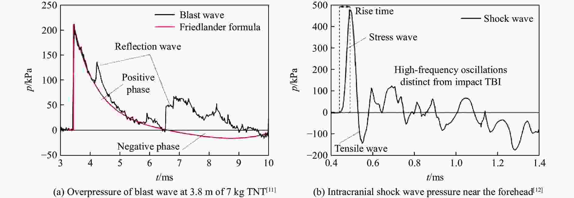
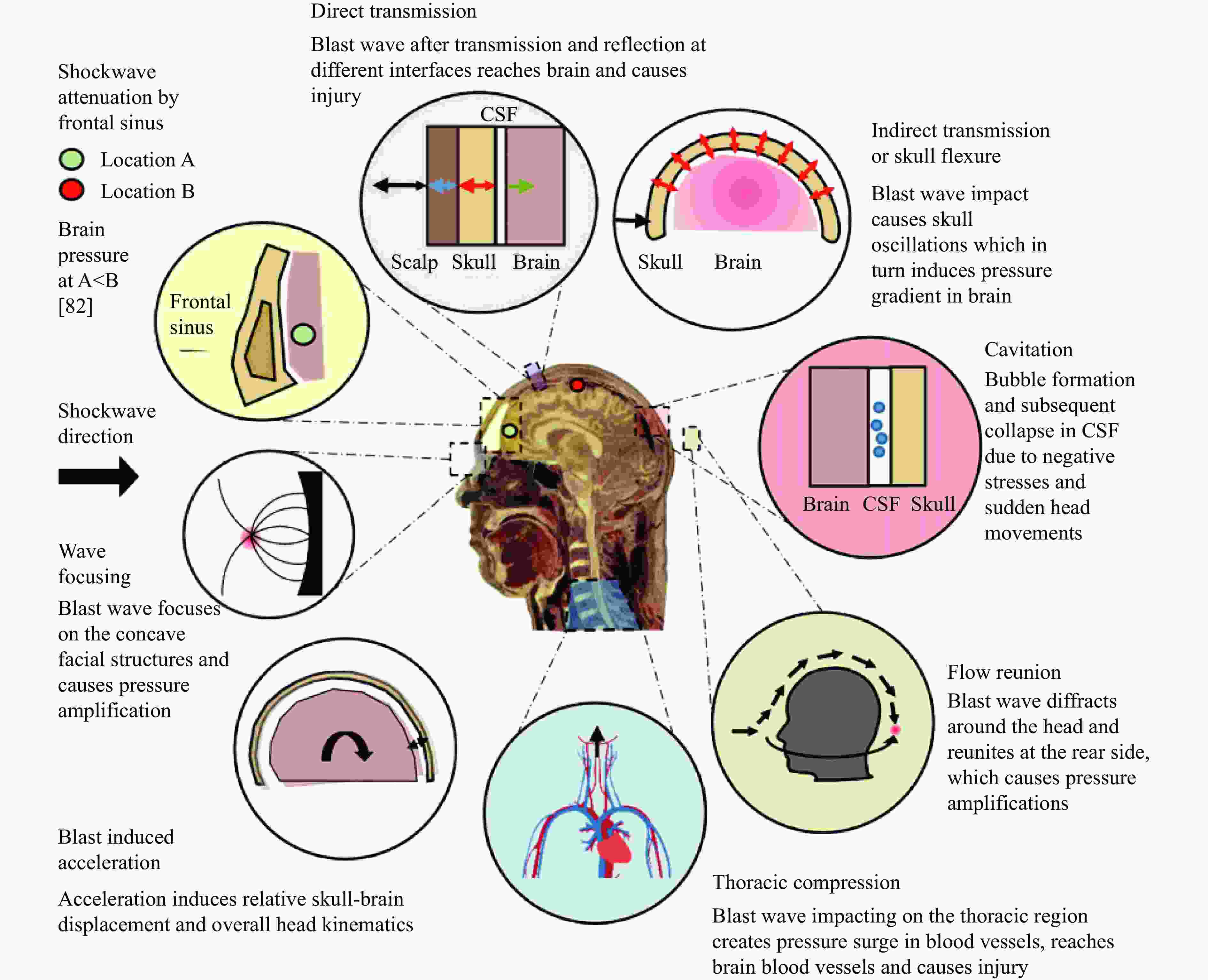
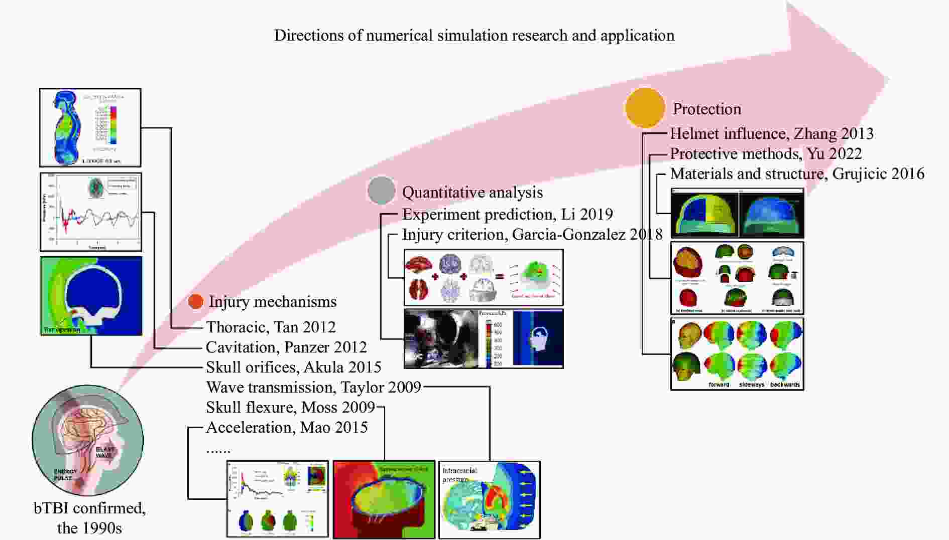
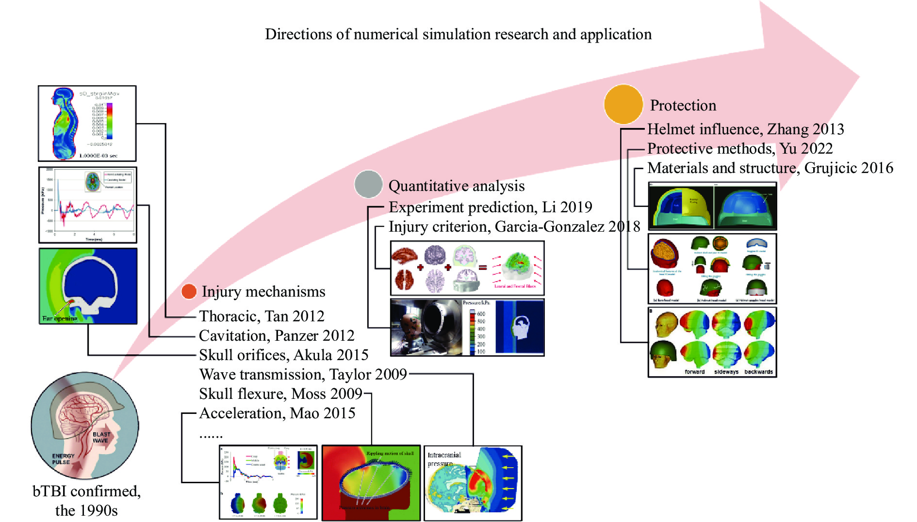
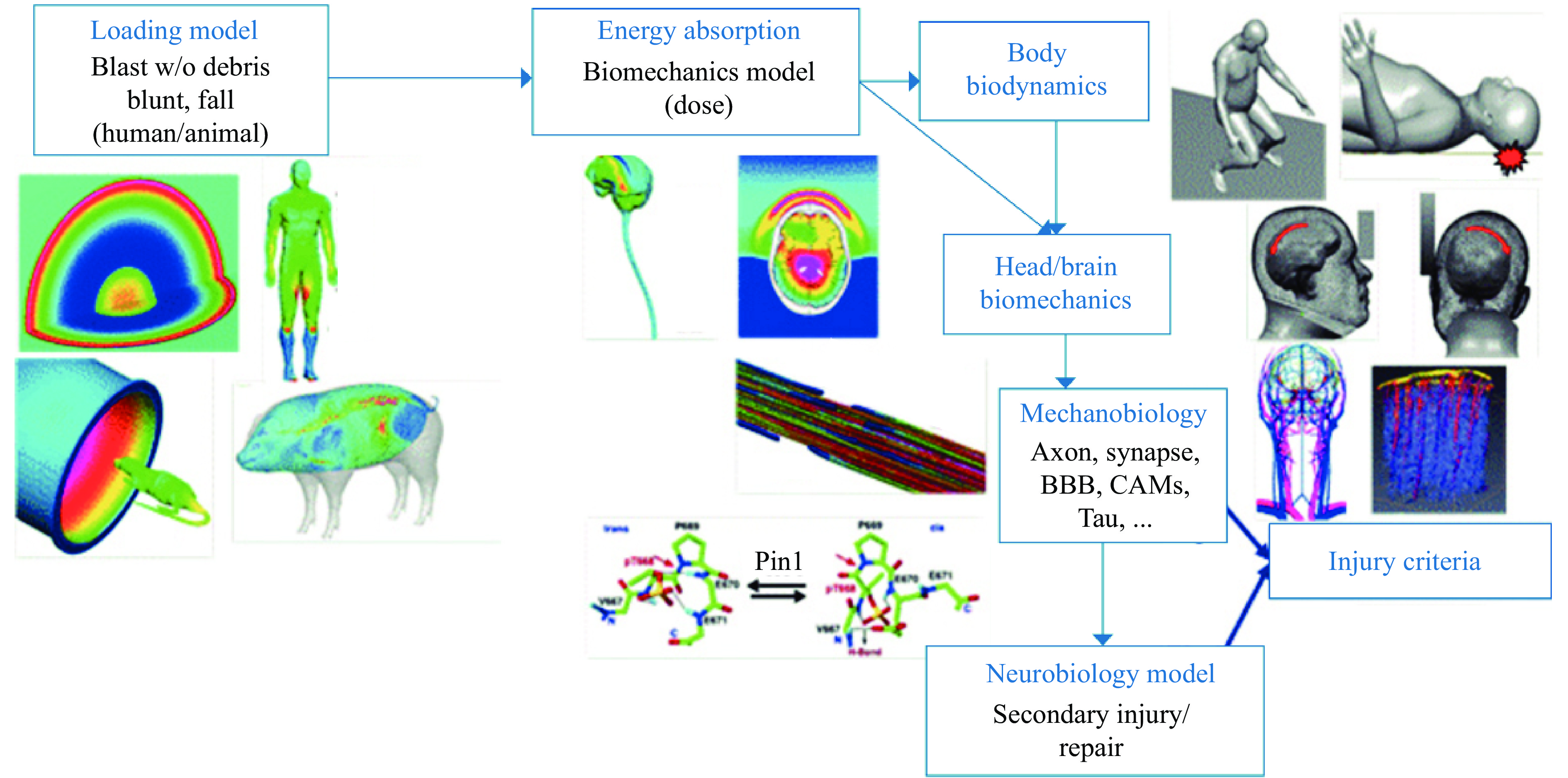
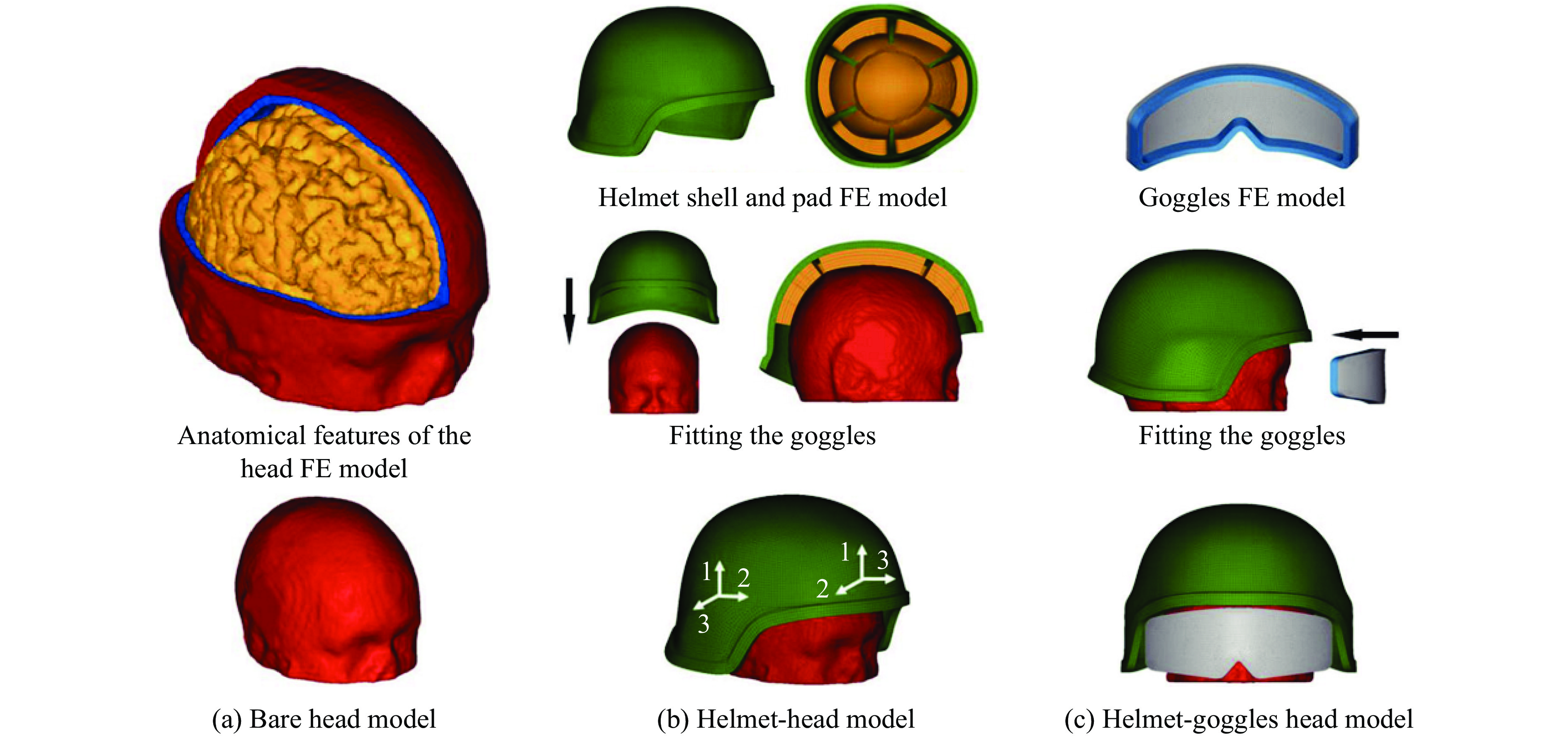
图 4 颅脑爆炸伤数值模拟研究与应用方向
Figure 4. Directions of numerical simulation research and application of bTBI


 下载: 导出CSV
下载: 导出CSV
下载: 导出CSV
表 3 脑组织超弹性本构模型分类
Table 3. Classification of hyper-elastic constitutive models of brain tissue
分类 模型名称 应变能函数 量符号解释 多项式形式 Neo-Hookean $ W = \dfrac{{{C_1}}}{2}\left( {{I_1} - 3} \right) $ C1为材料常数,I1为应变不变量 Mooney-Rivlin $ W = \displaystyle\sum\limits_{i + j = N}^N {{C_{ij}}{{\left( {{I_1} - 3} \right)}^i}{{\left( {{I_2} - 3} \right)}^j}} $ N为模型阶数,C1为材料常数,
I1、I2为应变不变量Ogden $ W = \displaystyle\sum\limits_{k = 1}^N {\dfrac{{{\mu _k}}}{{{\alpha _k}}}\left( {\lambda _1^{{\alpha _k}} + \lambda _2^{{\alpha _k}} + \lambda _3^{{\alpha _k}} - 3} \right)} $ μk和αk为材料常数,λ1、λ2、λ3为主拉伸比 Yeoh $ W = \displaystyle\sum\limits_{i = 1}^N {{C_i}{{\left( {{I_1} - 3} \right)}^i}} $ Ci为材料常数,I1为应变不变量 指数或
对数形式Fung-Demiray $ W = \dfrac{{{C_1}}}{{2{C_2}}}\left( {{{\text{e}}^{{C_2}\left( {{I_1} - 3} \right)}} - 1} \right) $ C1、C2为材料常数,I1为应变不变量 Veronda-Westmann $ W = {C_1}\left( {{{\text{e}}^{{C_3}\left( {{I_1} - 3} \right)}} - 1} \right){\text{ + }}{C_2}\left( {{I_2} - 3} \right) $ C1、C2、C3为材料常数,I1、I2为应变不变量 Gent $ W = {{ - }}\dfrac{{\mu {J_m}}}{2}\ln \left( {1 - \dfrac{{{I_1} - 3}}{{{J_m}}}} \right){\text{ }}{I_1} {\text{<}} {J_m} + 3 $ μ和Jm为材料常数,I1为应变不变量 混合形式 Chui $ W = {{ - }}\dfrac{{{C_1}}}{2}\ln \left[ {1 - {C_2}\left( {{I_1} - 3} \right) + {C_3}\left( {{I_1} - 3} \right)} \right] $ C1、C2、C3为材料常数,I1为应变不变量 Gao $ \begin{array}{l}W = {{ - }}{C_1}\ln \left[ {1 - {C_2}\left( {\lambda _1^{{\alpha _1}} + \lambda _2^{{\alpha _1}} + \lambda _3^{{\alpha _1}} - 3} \right)} \right] + {C_3}\left( {\lambda _1^{{\alpha _1}} + \lambda _2^{{\alpha _1}} + \lambda _3^{{\alpha _1}} - 3} \right) \\ W = {C_1}\left[ {{{\text{e}}^{{C_2}\left( {\lambda _1^{{\alpha _1}} + \lambda _2^{{\alpha _1}} + \lambda _3^{{\alpha _1}}} \right)}} - 1} \right] + {C_3}\left( {\lambda _1^{{\alpha _1}} + \lambda _2^{{\alpha _1}} + \lambda _3^{{\alpha _1}} - 3} \right) \end{array}$ C1、C2、C3、α1、α2、α3为材料常数,
λ1、λ2、λ3为主拉伸比
下载: 导出CSV
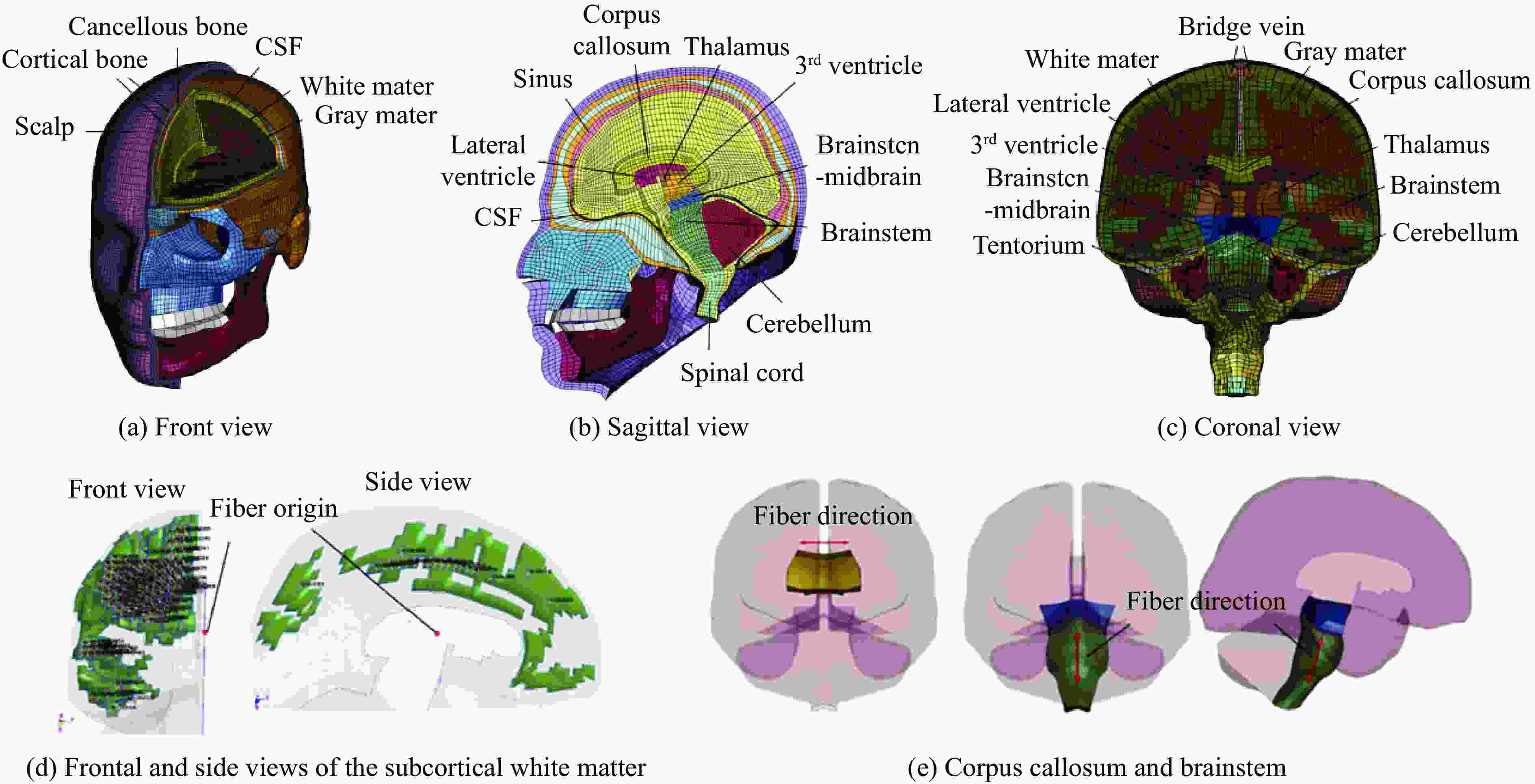
表 4 自2009年以来基于特征多块技术的bTBI模型
Table 4. Modeling of bTBI based on the feature multi-block technique since 2009
年份 模型来源 网格尺寸/mm 模型描述 模型验证 2009 文献[80-81] 流场:1
头部:1头部模型:美国国家医学图书馆-可视人体女性数据集
本构模型:颅骨(线弹性);白质和灰质(线性黏弹性);脑脊液(线弹性流体)尸体头部
撞击实验[82]2010
2016文献[47,83] 流场:不明确
头部:1~6头部模型:美国国立卫生研究院-可视人体数据库
本构模型:头皮、颅骨、硬脑膜、镰、小脑幕、软脑膜(线弹性);
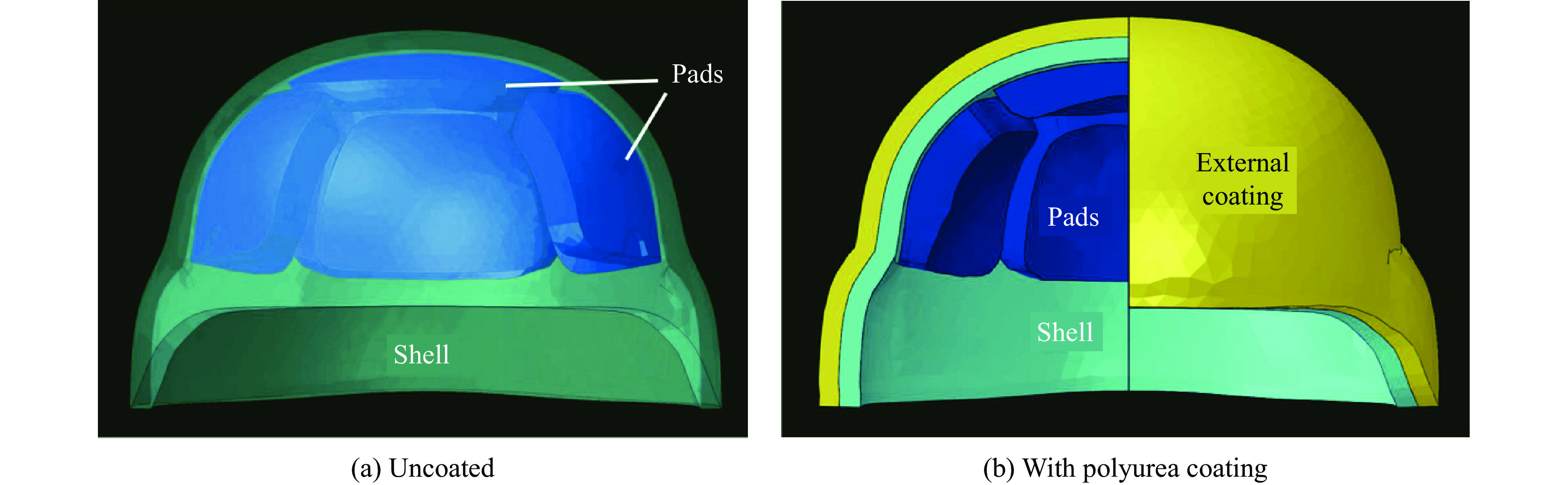
脑脊液(线弹性流体);大脑(黏超弹性)尸体头部
撞击实验[84]2011~2014 文献[85-87,31] 流场:10
头部:3~4头部模型:WSUHIM。
本构模型:面部(线弹性);灰质和白质、脑干、小脑(黏超弹性)尸体头部
激波管实验[85]2012 文献[88] 不明确 头部模型:美国国家交通研究中心-HSHM代理模型
本构模型:面部、颈部(线弹性);颅骨(线弹性、黏弹性);大脑(黏弹性)人头代理模型
激波管实验[88]2012 文献[48] 流场:30
头部:不明确本构模型:国际大脑测绘联盟-ICBM 2011数据库
本构模型:颅骨(线弹性);脑脊液(状态方程);大脑(线弹性与线性黏弹性);
桥静脉(超弹性)尸体头部
撞击实验[82]2014 文献[89] 流场:1
头部:1头部模型:美国国家医学图书馆-可视人体男性数据集
本构模型:颅骨、皮肤、椎骨(线弹性);肌肉/软组织(超弹性);
脑脊液(线弹性流体);大脑(线性黏弹性)尸体头部
激波管实验[90]2017 文献[91-92] 流场:7
头部:不明确头部模型:北达科他州立大学-NDSUHM模型
本构模型:头皮、面部骨骼和头骨、硬脑膜、镰、小脑幕、颈椎(线弹性);
大脑(黏超弹性);脑脊液(线弹性流体)尸体头部
撞击实验[82]2018 文献[93] 流场:10
头部:1头部模型:加拿大国防研究发展中心-代理模型
本构模型:颅骨、镰、小脑幕(线弹性);大脑(黏弹性)代理模型
爆炸实验[93]2019 文献[94,29] 流场:最小 3
头部:2~3头部模型:清华大学-充分反映颅脑生理结构
本构模型:大脑、小脑与脑干(黏超弹性);脑膜、蛛网膜、硬脑膜、大脑镰、小脑 幕、头骨、颈部与皮肤(线弹性);脑脊液(状态方程)尸体头部
撞击实验[82]2019~2020 文献[95-96] 流场:2.5
头部:不明确头部模型:帝国理工学院-反映脑沟生理结构
本构模型:颅骨、头皮、脑白质(线性黏弹性);脑脊液(状态方程)2021 文献[39] 流场:6
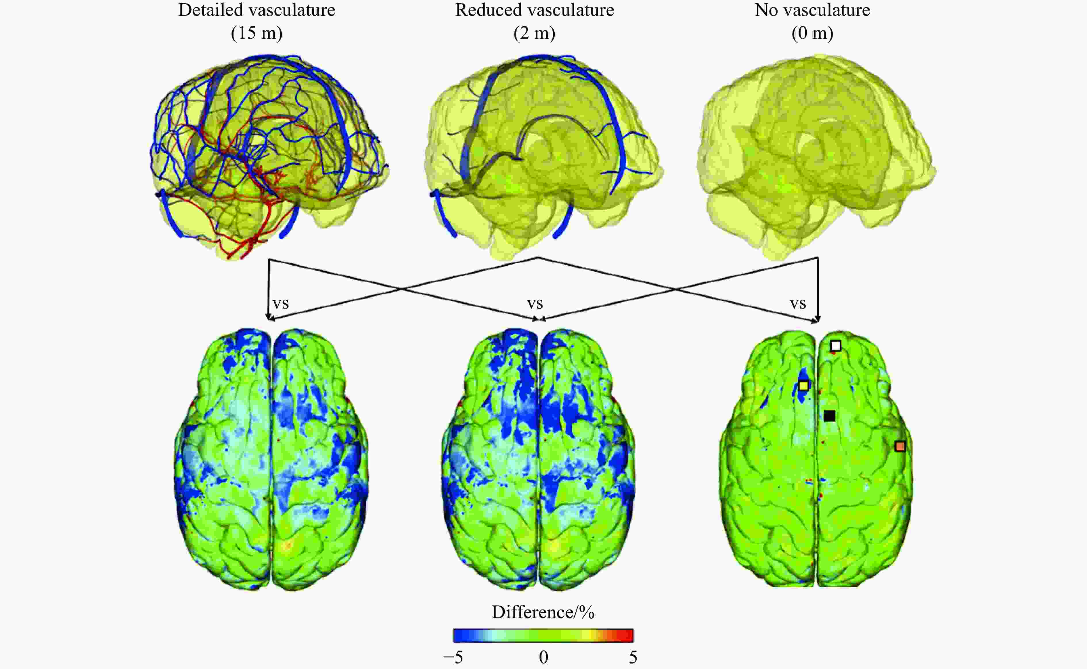
头部:0.1~2.3头部模型:美国男性50百分位模型-反映头部血管系统生理结构,总长度达15 m
本构模型:颅骨(线弹性);动脉、头皮、静脉、眼、脑膜(超弹性);大脑(黏超弹性)尸体头部
撞击实验[82,84,97]2022 文献[98] 流场:6
头部:最小
0.55头部模型:全球人体模型联盟- GHBMC v1.5
本构模型:白质和灰质、镰、小脑幕、胼胝体、面部、头皮等(黏超弹性);
脑膜、皮肤、颌、窦、桥静脉等(线弹性)尸体头部
激波管实验[90,99]
下载: 导出CSV
-
[1] 胡鹏伟, 张磊, 解宏伟, 等. 现代战争爆炸伤特点系统评价 [J]. 第二军医大学学报, 2021, 42(6): 681–687. DOI: 10.16781/j.0258-879x.2021.06.0681.HU P W, ZHANG L, XIE H W, et al. Characteristics of blast injuries in modern warfare: a systematic review [J]. Academic Journal of Second Military Medical University, 2021, 42(6): 681–687. DOI: 10.16781/j.0258-879x.2021.06.0681. [2] 柳占立, 杜智博, 张家瑞, 等. 颅脑爆炸伤致伤机制及防护研究进展 [J]. 爆炸与冲击, 2022, 42(4): 041101. DOI: 10.11883/bzycj-2021-0053.LIU Z L, DU Z B, ZHANG J R, et al. Progress in the mechanism and protection of blast-induced traumatic brain injury [J]. Explosion and Shock Waves, 2022, 42(4): 041101. DOI: 10.11883/bzycj-2021-0053. [3] 蔡志华, 贺葳, 汪剑辉, 等. 爆炸波致颅脑损伤力学机制与防护综述 [J]. 兵工学报, 2022, 43(2): 467–480. DOI: 10.3969/j.issn.1000-1093.2022.02.025.CAI Z H, HE W, WANG J H, et al. Review on mechanical mechanism of blast-induced traumatic brain injury and protection technology [J]. Acta Armamentarii, 2022, 43(2): 467–480. DOI: 10.3969/j.issn.1000-1093.2022.02.025. [4] MACLEOD A D. Shell shock, Gordon Holmes and the great war [J]. Journal of the Royal Society of Medicine, 2004, 97(2): 86–89. DOI: 10.1177/014107680409700215. [5] CERNAK I, WANG Z G, JIANG J X, et al. Ultrastructural and functional characteristics of blast injury-induced neurotrauma [J]. The Journal of Trauma: Injury, Infection, and Critical Care, 2001, 50(4): 695–706. DOI: 10.1097/00005373-200104000-00017. [6] CERNAK I, NOBLE-HAEUSSLEIN L J. Traumatic brain injury: an overview of pathobiology with emphasis on military populations [J]. Journal of Cerebral Blood Flow and Metabolism, 2010, 30(2): 255–266. DOI: 10.1038/jcbfm.2009.203. [7] BHATTACHARJEE Y. Shell shock revisited: solving the puzzle of blast trauma [J]. Science, 2008, 319(5862): 406–408. DOI: 10.1126/science.319.5862.406. [8] DEPALMA R G, HOFFMAN S W. Combat blast related traumatic brain injury (TBI): decade of recognition; promise of progress [J]. Behavioural Brain Research, 2018, 340: 102–105. DOI: 10.1016/j.bbr.2016.08.036. [9] 宁亚蕾, 周元国. 原发性颅脑冲击伤致伤机制及病理学特点 [J]. 中华创伤杂志, 2014, 30(3): 280–283. DOI: 10.3760/cma.j.issn.1001-8050.2014.03.024.NING Y L, ZHOU Y G. Pathogenesis and pathological characteristics of primary traumatic brain injury [J]. Chinese Journal of Trauma, 2014, 30(3): 280–283. DOI: 10.3760/cma.j.issn.1001-8050.2014.03.024. [10] TANIELIAN T, JAYCOX L H, SCHELL T L, et al. Invisible wounds of war: summary and recommendations for addressing psychological and cognitive injuries [M]. Santa Monica: RAND Corporation, 2008. DOI: 10.7249/MG720.1. [11] LI J T, MA T, HUANG C, et al. Protective mechanism of helmet under far-field shock wave [J]. International Journal of Impact Engineering, 2020, 143: 103617. DOI: 10.1016/j.ijimpeng.2020.103617. [12] 栗志杰, 由小川, 柳占立, 等. 爆炸冲击波作用下颅脑损伤机理的数值模拟研究 [J]. 爆炸与冲击, 2020, 40(1): 015901. DOI: 10.11883/bzycj-2018-0348.LI Z J, YOU X C, LIU Z L, et al. Numerical simulation of the mechanism of traumatic brain injury induced by blast shock waves [J]. Explosion and Shock Waves, 2020, 40(1): 015901. DOI: 10.11883/bzycj-2018-0348. [13] 赵辉, 朱峰. 原发性颅脑冲击伤的生物力学机制 [J]. 创伤外科杂志, 2016, 18(6): 375–378. DOI: 10.3969/j.issn.1009-4237.2016.06.017.ZHAO H, ZHU F. The biomechanical mechanism of primary blast brain injury [J]. Journal of Traumatic Surgery, 2016, 18(6): 375–378. DOI: 10.3969/j.issn.1009-4237.2016.06.017. [14] ZHU F, CHOU C C, YANG K H, et al. A theoretical analysis of stress wave propagation in the head under primary blast loading [J]. Proceedings of the Institution of Mechanical Engineers, Part H: Journal of Engineering in Medicine, 2014, 228(5): 439–445. DOI: 10.1177/0954411914530882. [15] COURTNEY A, COURTNEY M. The complexity of biomechanics causing primary blast-induced traumatic brain injury: a review of potential mechanisms [J]. Frontiers in Neurology, 2015, 6: 221. DOI: 10.3389/fneur.2015.00221. [16] ALLEY M D, SCHIMIZZE B R, SON S F. Experimental modeling of explosive blast-related traumatic brain injuries [J]. NeuroImage, 2011, 54: S45–S54. DOI: 10.1016/j.neuroimage.2010.05.030. [17] MOSS W C, KING M J, BLACKMAN E G. Skull flexure from blast waves: a mechanism for brain injury with implications for helmet design [J]. Physical Review Letters, 2009, 103(10): 108702. DOI: 10.1103/physrevlett.103.108702. [18] BOLANDER R, MATHIE B, BIR C, et al. Skull flexure as a contributing factor in the mechanism of injury in the rat when exposed to a shock wave [J]. Annals of Biomedical Engineering, 2011, 39(10): 2550–2559. DOI: 10.1007/s10439-011-0343-0. [19] GARIMELLA H T, KRAFT R H, PRZEKWAS A J. Do blast induced skull flexures result in axonal deformation? [J]. PLoS One, 2018, 13(3): e0190881. DOI: 10.1371/journal.pone.0190881. [20] HONG Y, SARNTINORANONT M, SUBHASH G, et al. Localized tissue surrogate deformation due to controlled single bubble cavitation [J]. Experimental Mechanics, 2016, 56(1): 97–109. DOI: 10.1007/s11340-015-0024-2. [21] CANCHI S, KELLY K, HONG Y, et al. Controlled single bubble cavitation collapse results in jet-induced injury in brain tissue [J]. Journal of the Mechanical Behavior of Biomedical Materials, 2017, 74: 261–273. DOI: 10.1016/j.jmbbm.2017.06.018. [22] CHEN Y, HUANG W. Non-impact, blast-induced mild TBI and PTSD: concepts and caveats [J]. Brain Injury, 2011, 25(7/8): 641–650. DOI: 10.3109/02699052.2011.580313. [23] SUNDAR S, PONNALAGU A. Biomechanical analysis of head subjected to blast waves and the role of combat protective headgear under blast loading: a review [J]. Journal of Biomechanical Engineering, 2021, 143(10): 100801. DOI: 10.1115/1.4051047. [24] AKULA P, HUA Y, GU L X. Blast-induced mild traumatic brain injury through ear canal: a finite element study [J]. Biomedical Engineering Letters, 2015, 5(4): 281–288. DOI: 10.1007/s13534-015-0204-0. [25] TAYLOR P A, FORD C C. Simulation of blast-induced early-time intracranial wave physics leading to traumatic brain injury [J]. Journal of Biomechanical Engineering, 2009, 131(6): 061007. DOI: 10.1115/1.3118765. [26] PANZER M B, MYERS B S, CAPEHART B P, et al. Development of a finite element model for blast brain injury and the effects of CSF cavitation [J]. Annals of Biomedical Engineering, 2012, 40(7): 1530–1544. DOI: 10.1007/s10439-012-0519-2. [27] TAN X G, KANNAN R, PRZEKWAS A J. A comparative study of the human body finite element model under blast loadings [C]//ASME 2012 International Mechanical Engineering Congress and Exposition. Houston, USA: American Society of Mechanical Engineers, 2012: 829–836. DOI: 10.1115/IMECE2012-89072. [28] MAO H J, UNNIKRISHNAN G, RAKESH V, et al. Untangling the effect of head acceleration on brain responses to blast waves [J]. Journal of Biomechanical Engineering, 2015, 137(12): 124502. DOI: 10.1115/1.4031765. [29] LI Z J, DU Z B, YOU X C, et al. Numerical study on dynamic mechanism of brain volume and shear deformation under blast loading [J]. Acta Mechanica Sinica, 2019, 35(5): 1104–1119. DOI: 10.1007/s10409-019-00875-w. [30] GARCIA-GONZALEZ D, RACE N S, VOETS N L, et al. Cognition based bTBI mechanistic criteria: a tool for preventive and therapeutic innovations [J]. Scientific Reports, 2018, 8(1): 10273. DOI: 10.1038/s41598-018-28271-7. [31] ZHANG L Y, MAKWANA R, SHARMA S. Brain response to primary blast wave using validated finite element models of human head and advanced combat helmet [J]. Frontiers in Neurology, 2013, 4: 88. DOI: 10.3389/fneur.2013.00088. [32] YU X C, GHAJARI M. Protective performance of helmets and goggles in mitigating brain biomechanical response to primary blast exposure [J]. Annals of Biomedical Engineering, 2022, 50(11): 1579–1595. DOI: 10.1007/s10439-022-02936-x. [33] GRUJICIC M, RAMASWAMI S, SNIPES J S, et al. Experimental and computational investigations of the potential improvement in helmet blast-protection through the use of a polyurea-based external coating [J]. Multidiscipline Modeling in Materials and Structures, 2016, 12(1): 33–72. DOI: 10.1108/MMMS-02-2015-0009. [34] HOLBOURN A H S. Mechanics of head injuries [J]. The Lancet, 1943, 242(6267): 438–441. DOI: 10.1016/S0140-6736(00)87453-X. [35] SHUGAR T A, KATONA M G. Development of finite element head injury model [J]. Journal of the Engineering Mechanics Division, 1975, 101(3): 223–239. DOI: 10.1061/JMCEA3.0002012. [36] SHUGAR T A. A finite element head injury model: DOT HS 803-211 [R]. Washington DC, USA: Department of Transportation, National Highway Traffic Safety Administration, 1977. [37] WRIGHT R M, POST A, HOSHIZAKI B, et al. A multiscale computational approach to estimating axonal damage under inertial loading of the head [J]. Journal of Neurotrauma, 2013, 30(2): 102–118. DOI: 10.1089/neu.2012.2418. [38] BILSTON L E. Neural tissue biomechanics: Vol 3 [M]. Berlin, Germany: Springer, 2011. DOI: 10.1007/978-3-642-13890-4. [39] SUBRAMANIAM D R, UNNIKRISHNAN G, SUNDARAMURTHY A, et al. Cerebral vasculature influences blast-induced biomechanical responses of human brain tissue [J]. Frontiers in Bioengineering and Biotechnology, 2021, 9: 744808. DOI: 10.3389/fbioe.2021.744808. [40] BASTIANI M, ROEBROECK A. Unraveling the multiscale structural organization and connectivity of the human brain: the role of diffusion MRI [J]. Frontiers in Neuroanatomy, 2015, 9: 77. DOI: 10.3389/fnana.2015.00077. [41] PATEL N, KIRMI O. Anatomy and imaging of the normal meninges [J]. Seminars in Ultrasound, CT and MRI, 2009, 30(6): 559–564. DOI: 10.1053/j.sult.2009.08.006. [42] SNELL R S. Clinical neuroanatomy [M]. 7th ed. Philadelphia, PA: Wolters Kluwer Health/Lippincott Williams and Wilkins, 2010. [43] SAAB A S, NAVE K A. Myelin dynamics: protecting and shaping neuronal functions [J]. Current Opinion in Neurobiology, 2017, 47: 104–112. DOI: 10.1016/j.conb.2017.09.013. [44] PATESTAS M A, GARTNER L P. A textbook of neuroanatomy [M]. 2nd ed. Hoboken, New Jersey, USA: John Wiley & Sons, Inc. , 2016. [45] MONTANINO A. Definition of axonal injury tolerances across scales: a computational multiscale approach [D]. Stockholm: Kungliga Tekniska Högskolan, 2020: 6-10. [46] GANPULE S, ALAI A, PLOUGONVEN E, et al. Mechanics of blast loading on the head models in the study of traumatic brain injury using experimental and computational approaches [J]. Biomechanics and Modeling in Mechanobiology, 2013, 12(3): 511–531. DOI: 10.1007/s10237-012-0421-8. [47] WANG C Z, PAHK J B, BALABAN C D, et al. Biomechanical assessment of the bridging vein rupture of blast-induced traumatic brain injury using the finite element human head model [C]//ASME 2012 International Mechanical Engineering Congress and Exposition. Houston, USA: American Society of Mechanical Engineers, 2012: 795−805. DOI: 10.1115/IMECE2012-88739. [48] MOORE D F, JÉRUSALEM A, NYEIN M, et al. Computational biology: modeling of primary blast effects on the central nervous system [J]. NeuroImage, 2009, 47(Suppl 2): 10–20. DOI: 10.1016/j.neuroimage.2009.02.019. [49] CHAFI M S, KARAMI G, ZIEJEWSKI M. Biomechanical assessment of brain dynamic responses due to blast pressure waves [J]. Annals of Biomedical Engineering, 2010, 38(2): 490–504. DOI: 10.1007/s10439-009-9813-z. [50] TAKHOUNTS E G, RIDELLA S A, HASIJA V, et al. Investigation of traumatic brain injuries using the next generation of simulated injury monitor (SIMon) finite element head model [J]. Stapp Car Crash Journal, 2008, 52: 1–31. DOI: 10.4271/2008-22-0001. [51] VIANO D C, CASSON I R, PELLMAN E J, et al. Concussion in professional football: brain responses by finite element analysis: Part 9 [J]. Neurosurgery, 2005, 57(5): 891–916. DOI: 10.1227/01.NEU.0000186950.54075.3B. [52] MENDIS K K, STALNAKER R L, ADVANI S H. A constitutive relationship for large deformation finite element modeling of brain tissue [J]. Journal of Biomechanical Engineering, 1995, 117(3): 279–285. DOI: 10.1115/1.2794182. [53] MILLER K, CHINZEI K, ORSSENGO G, et al. Mechanical properties of brain tissue in-vivo: experiment and computer simulation [J]. Journal of Biomechanics, 2000, 33(11): 1369–1376. DOI: 10.1016/S0021-9290(00)00120-2. [54] RASHID B, DESTRADE M, GILCHRIST M D. Mechanical characterization of brain tissue in simple shear at dynamic strain rates [J]. Journal of the Mechanical Behavior of Biomedical Materials, 2013, 28: 71–85. DOI: 10.1016/j.jmbbm.2013.07.017. [55] WEX C, ARNDT S, STOLL A, et al. Isotropic incompressible hyperelastic models for modelling the mechanical behaviour of biological tissues: a review [J]. Biomedical Engineering, 2015, 60(6): 577–592. DOI: 10.1515/bmt-2014-0146. [56] LAKSARI K, SHAFIEIAN M, DARVISH K. Constitutive model for brain tissue under finite compression [J]. Journal of Biomechanics, 2012, 45(4): 642–646. DOI: 10.1016/j.jbiomech.2011.12.023. [57] OGDEN R W. Large deformation isotropic elasticity: on the correlation of theory and experiment for incompressible rubberlike solids [J]. Proceedings of the Royal Society A: Mathematical, Physical and Engineering Sciences, 1972, 326(1567): 565–584. [58] KOHANDEL M, SIVALOGANATHAN S, TENTI G, et al. The constitutive properties of the brain parenchyma. Part 1: strain energy approach [J]. Medical Engineering and Physics, 2006, 28(5): 449–454. DOI: 10.1016/j.medengphy.2005.01.005. [59] KASTER T, SACK I, SAMANI A. Measurement of the hyperelastic properties of ex vivo brain tissue slices [J]. Journal of Biomechanics, 2011, 44(6): 1158–1163. DOI: 10.1016/j.jbiomech.2011.01.019. [60] COATS B, MARGULIES S S. Material properties of porcine parietal cortex [J]. Journal of Biomechanics, 2006, 39(13): 2521–2525. DOI: 10.1016/j.jbiomech.2005.07.020. [61] RASHID B, DESTRADE M, GILCHRIST M D. Inhomogeneous deformation of brain tissue during tension tests [J]. Computational Materials Science, 2012, 64: 295–300. DOI: 10.1016/j.commatsci.2012.05.030. [62] RASHID B, DESTRADE M, GILCHRIST M D. Mechanical characterization of brain tissue in tension at dynamic strain rates [J]. Journal of the Mechanical Behavior of Biomedical Materials, 2014, 33: 43–54. DOI: 10.1016/j.jmbbm.2012.07.015. [63] SPARREY C J, KEAVENY T M. Compression behavior of porcine spinal cord white matter [J]. Journal of Biomechanics, 2011, 44(6): 1078–1082. DOI: 10.1016/j.jbiomech.2011.01.035. [64] O’HAGAN J J, SAMANI A. Measurement of the hyperelastic properties of 44 pathological ex vivo breast tissue samples [J]. Physics in Medicine and Biology, 2009, 54(8): 2557–2569. DOI: 10.1088/0031-9155/54/8/020. [65] KLINICH K D, MILLER C S, HU J, et al. Characterization of ovine utero-placental interface tensile failure [J]. Placenta, 2012, 33(10): 776–781. DOI: 10.1016/j.placenta.2012.06.012. [66] CHUI C, KOBAYASHI E, CHEN X, et al. Combined compression and elongation experiments and non-linear modelling of liver tissue for surgical simulation [J]. Medical and Biological Engineering and Computing, 2004, 42(6): 787–798. DOI: 10.1007/BF02345212. [67] 路纯红, 白鸿柏. 粘弹性材料本构模型的研究 [J]. 高分子材料科学与工程, 2007, 23(6): 28–31, 35. DOI: 10.3321/j.issn:1000-7555.2007.06.007.LU C H, BAI H B. Study on constitutive model of viscoelastic material [J]. Polymer Materials Science and Engineering, 2007, 23(6): 28–31, 35. DOI: 10.3321/j.issn:1000-7555.2007.06.007. [68] PRABHU R, HORSTEMEYER M F, TUCKER M T, et al. Coupled experiment/finite element analysis on the mechanical response of porcine brain under high strain rates [J]. Journal of the Mechanical Behavior of Biomedical Materials, 2011, 4(7): 1067–1080. DOI: 10.1016/j.jmbbm.2011.03.015. [69] COLGAN N C, GILCHRIST M D, CURRAN K M. Applying DTI white matter orientations to finite element head models to examine diffuse TBI under high rotational accelerations [J]. Progress in Biophysics and Molecular Biology, 2010, 103(2/3): 304–309. DOI: 10.1016/j.pbiomolbio.2010.09.008. [70] CHATELIN S, DECK C, RENARD F, et al. Computation of axonal elongation in head trauma finite element simulation [J]. Journal of the Mechanical Behavior of Biomedical Materials, 2011, 4(8): 1905–1919. DOI: 10.1016/j.jmbbm.2011.06.007. [71] CLOOTS R J H, VAN DOMMELEN J A W, KLEIVEN S, et al. Multi-scale mechanics of traumatic brain injury: predicting axonal strains from head loads [J]. Biomechanics and Modeling in Mechanobiology, 2013, 12(1): 137–150. DOI: 10.1007/s10237-012-0387-6. [72] SAHOO D, DECK C, WILLINGER R. Development and validation of an advanced anisotropic visco-hyperelastic human brain FE model [J]. Journal of the Mechanical Behavior of Biomedical Materials, 2014, 33: 24–42. DOI: 10.1016/j.jmbbm.2013.08.022. [73] GIORDANO C, CLOOTS R J H, VAN DOMMELEN J A W, et al. The influence of anisotropy on brain injury prediction [J]. Journal of Biomechanics, 2014, 47(5): 1052–1059. DOI: 10.1016/j.jbiomech.2013.12.036. [74] GIORDANO C, ZAPPALÀ S, KLEIVEN S. Anisotropic finite element models for brain injury prediction: the sensitivity of axonal strain to white matter tract inter-subject variability [J]. Biomechanics and Modeling in Mechanobiology, 2017, 16(4): 1269–1293. DOI: 10.1007/s10237-017-0887-5. [75] BAECK K, GOFFIN J, VANDER SLOTEN J. The use of different CSF representations in a numerical head model and their effect on the results of FE head impact analyses [C]//8th European LS-DYNA Users Conference. Strasbourg: DYNAmore, 2011. [76] BREWICK P, TEFERRA K. Addressing uncertainty in constitutive model forms and parameters for FE models of the human head subjected to blast loading [C]//ASME 2017 International Mechanical Engineering Congress and Exposition. Tampa: American Society of Mechanical Engineers, 2017: V003T04A032. DOI: 10.1115/IMECE2017-70281. [77] GIUDICE J S, ZENG W, WU T T, et al. An analytical review of the numerical methods used for finite element modeling of traumatic brain injury [J]. Annals of Biomedical Engineering, 2019, 47(9): 1855–1872. DOI: 10.1007/s10439-018-02161-5. [78] TSE K M, LIM S P, TAN V B C, et al. A review of head injury and finite element head models [J]. American Journal of Engineering, Technology and Society, 2014, 1(5): 28–52. [79] DIXIT P, LIU G R. A review on recent development of finite element models for head injury simulations [J]. Archives of Computational Methods in Engineering, 2017, 24(4): 979–1031. DOI: 10.1007/s11831-016-9196-x. [80] TAYLOR P A, FORD C C. Simulation and correlation of blast-induced early-time intracranial wave physics with traumatic brain injury: SAND2010-0371C [R]. Albuquerque, USA: Sandia National Laboratories, 2010. [81] TAYLOR P A, LUDWIGSEN J S, FORD C C. Investigation of blast-induced traumatic brain injury [J]. Brain Injury, 2014, 28(7): 879–895. DOI: 10.3109/02699052.2014.888478. [82] NAHUM A M, SMITH R, WARD C C. Intracranial pressure dynamics during head impact [C]//SAE Technical Papers. USA: SAE International, 1977. DOI: 10.4271/770922. [83] JAZI M S, REZAEI A, AZARMI F, et al. Computational biomechanics of human brain with and without the inclusion of the body under different blast orientation [J]. Computer Methods in Biomechanics and Biomedical Engineering, 2016, 19(9): 1019–1031. DOI: 10.1080/10255842.2015.1088525. [84] HARDY W N, MASON M J, FOSTER C D, et al. A study of the response of the human cadaver head to impact [J]. Stapp Car Crash Journal, 2007, 51: 17–80. DOI: 10.4271/2007-22-0002. [85] SHARMA S. Biomechanical analysis of blast induced traumatic brain injury: a finite element modeling and validation study of blast effects on human brain [D]. Detroit, Michigan, USA: Wayne State University, 2011. [86] MAKWANA R. Development and validation of a three-dimensional finite element model of advanced combat helmet and biomechanical analysis of human head and helmet response to primary blast insult [D]. Detroit, Michigan, USA: Wayne State University, 2012. [87] MAKWANA R, SHARMA S, ZHANG L Y. Comparison of the brain response to blast exposure between a human head model and a blast headform model using finite element methods [C]//13th International LS-DYNA Users Conference. Dearborn, Michigan, USA, 2014. [88] ROBERTS J C, HARRIGAN T P, WARD E E, et al. Human head-neck computational model for assessing blast injury [J]. Journal of Biomechanics, 2012, 45(16): 2899–2906. DOI: 10.1016/j.jbiomech.2012.07.027. [89] SINGH D, CRONIN D S, HALADUICK T N. Head and brain response to blast using sagittal and transverse finite element models [J]. International Journal for Numerical Methods in Biomedical Engineering, 2014, 30(4): 470–489. DOI: 10.1002/cnm.2612. [90] BIR C. Measuring blast-related intracranial pressure within the human head: W81XWH-09-1-0498 [R]. Fort Belvoir, VA: Defense Technical Information Center, 2011. DOI: 10.21236/ADA547306. [91] SARVGHAD-MOGHADDAM H, REZAEI A, ZIEJEWSKI M, et al. Correlative analysis of head kinematics and brain’s tissue response: a computational approach toward understanding the mechanisms of blast TBI [J]. Shock Waves, 2017, 27(6): 919–927. DOI: 10.1007/s00193-017-0749-1. [92] SARVGHAD-MOGHADDAM H, REZAEI A, ZIEJEWSKI M, et al. Evaluation of brain tissue responses because of the underwash overpressure of helmet and faceshield under blast loading [J]. International Journal for Numerical Methods in Biomedical Engineering, 2017, 33(1): e02782. DOI: 10.1002/cnm.2782. [93] DOWNES D, BOUAMOUL A, OUELLET S, et al. Development and validation of a biofidelic head form model to assess blast-induced traumatic brain injury [J]. The Journal of Defense Modeling and Simulation: Applications, Methodology, Technology, 2018, 15(3): 257–267. DOI: 10.1177/1548512917737634. [94] 栗志杰, 由小川, 柳占立, 等. 一种物理头部模型和测试系统: CN108168827B [P]. 2018-06-15. [95] YU X C, GHAJARI M. An assessment of blast modelling techniques for injury biomechanics research [J]. International Journal for Numerical Methods in Biomedical Engineering, 2019, 35(12): e3258. DOI: 10.1002/cnm.3258. [96] YU X C, AZOR A, J SHARP D, et al. Mechanisms of tensile failure of cerebrospinal fluid in blast traumatic brain injury [J]. Extreme Mechanics Letters, 2020, 38: 100739. DOI: 10.1016/j.eml.2020.100739. [97] TROSSEILLE X, TARRIÉRE C, LAVASTE F, et al. Development of a F. E. M. of the human head according to a specific test protocol [C]//Proceeding of the 36th Stapp Car Crash Conference. USA: SAE International, 1992. DOI: 10.4271/922527. [98] SUTAR S, GANPULE S. Evaluation of blast simulation methods for modeling blast wave interaction with human head [J]. Journal of Biomechanical Engineering, 2022, 144(5): 051009. DOI: 10.1115/1.4053059. [99] SKOTAK M, ALAY E, ZHENG J Q, et al. Effective testing of personal protective equipment in blast loading conditions in shock tube: comparison of three different testing locations [J]. PLoS One, 2018, 13(6): e0198968. DOI: 10.1371/journal.pone.0198968. [100] MAO H J, ZHANG L Y, JIANG B H, et al. Development of a finite element human head model partially validated with thirty five experimental cases [J]. Journal of Biomechanical Engineering, 2013, 135(11): 111002. DOI: 10.1115/1.4025101. [101] LYU D, ZHOU R Z, LIN C H, et al. Development and validation of a new anisotropic visco-hyperelastic human head finite element model capable of predicting multiple brain injuries [J]. Frontiers in Bioengineering and Biotechnology, 2022, 10: 831595. DOI: 10.3389/fbioe.2022.831595. [102] ABOLFATHI N, NAIK A, CHAFI M S, et al. A micromechanical procedure for modelling the anisotropic mechanical properties of brain white matter [J]. Computer Methods in Biomechanics and Biomedical Engineering, 2009, 12(3): 249–262. DOI: 10.1080/10255840802430587. [103] GRUJICIC M, D’ENTREMONT B, PANDURANGAN B, et al. A study of the blast-induced brain white-matter damage and the associated diffuse axonal injury [J]. Multidiscipline Modeling in Materials and Structures, 2012, 8(2): 213–245. DOI: 10.1108/15736101211251220. [104] JAVID S, REZAEI A, KARAMI G. A micromechanical procedure for viscoelastic characterization of the axons and ECM of the brainstem [J]. Journal of the Mechanical Behavior of Biomedical Materials, 2014, 30: 290–299. DOI: 10.1016/j.jmbbm.2013.11.010. [105] YOUSEFSANI S A, FARAHMAND F, SHAMLOO A. A three-dimensional micromechanical model of brain white matter with histology-informed probabilistic distribution of axonal fibers [J]. Journal of the Mechanical Behavior of Biomedical Materials, 2018, 88: 288–295. DOI: 10.1016/j.jmbbm.2018.08.042. [106] WU T T, ALSHAREEF A, GIUDICE J S, et al. Explicit modeling of white matter axonal fiber tracts in a finite element brain model [J]. Annals of Biomedical Engineering, 2019, 47(9): 1908–1922. DOI: 10.1007/s10439-019-02239-8. [107] GARIMELLA H T, KRAFT R H. Modeling the mechanics of axonal fiber tracts using the embedded finite element method [J]. International Journal for Numerical Methods in Biomedical Engineering, 2017, 33(5): e2823. DOI: 10.1002/cnm.2823. [108] GUPTA R K, PRZEKWAS A J. A framework for multiscale modeling of warfighter blast injury protection [C]//The 6th International Conference on Computational Methods (ICCM2015). Auckland, New Zealand: ScienTech Publisher, 2015: 7. [109] GUPTA R K, TAN X G, SOMAYAJI M R, et al. Multiscale modelling of blast-induced TBI mechanobiology: from body to neuron to molecule [J]. Defence Life Science Journal, 2017, 2(1): 3–13. DOI: 10.14429/dlsj.2.10369. [110] PRZEKWAS A, GARIMELLA H T, TAN X G, et al. Biomechanics of blast TBI with time-resolved consecutive primary, secondary, and tertiary loads [J]. Military Medicine, 2019, 184(S1): 195–205. DOI: 10.1093/milmed/usy344. [111] SUTAR S, GANPULE S. Investigation of wave propagation through head layers with focus on understanding blast wave transmission [J]. Biomechanics and Modeling in Mechanobiology, 2020, 19(3): 875–892. DOI: 10.1007/s10237-019-01256-9. [112] RUBIO J E, UNNIKRISHNAN G, SAJJA V S S S, et al. Investigation of the direct and indirect mechanisms of primary blast insult to the brain [J]. Scientific Reports, 2021, 11(1): 16040. DOI: 10.1038/s41598-021-95003-9. [113] NYEIN M K, JASON A M, YU L, et al. Reply to Moss et al: military and medically relevant models of blast-induced traumatic brain injury vs. ellipsoidal heads and helmets [J]. Proceedings of the National Academy of Sciences of the United States of America, 2011, 108(17): E83. DOI: 10.1073/pnas.1102626108. [114] GUPTA R K, PRZEKWAS A. Mathematical models of blast-induced TBI: current status, challenges, and prospects [J]. Frontiers in Neurology, 2013, 4: 59. DOI: 10.3389/fneur.2013.00059. [115] DU Z B, LI Z J, WANG P, et al. Revealing the effect of skull deformation on intracranial pressure variation during the direct interaction between blast wave and surrogate head [J]. Annals of Biomedical Engineering, 2022, 50(9): 1038–1052. DOI: 10.1007/s10439-022-02982-5. [116] UNNIKRISHNAN G, MAO H J, SUNDARAMURTHY A, et al. A 3-D rat brain model for blast-wave exposure: effects of brain vasculature and material properties [J]. Annals of Biomedical Engineering, 2019, 47(9): 2033–2044. DOI: 10.1007/s10439-019-02277-2. [117] 康越, 张仕忠, 张远平, 等. 基于激波管评价的单兵头面部装备冲击波防护性能研究 [J]. 爆炸与冲击, 2021, 41(8): 085901. DOI: 10.11883/bzycj-2020-0395.KANG Y, ZHANG S Z, ZHANG Y P, et al. Research on anti-shockwave performance of the protective equipment for the head of a soldier based on shock tube evaluation [J]. Explosion and Shock Waves, 2021, 41(8): 085901. DOI: 10.11883/bzycj-2020-0395. [118] RADOVITZKY R, SOCRATE S, TABER K, et al. Investigations of tissue-level mechanisms of primary blast injury through modeling, simulation, neuroimaging and neuropathological studies [R]. Fort Belvoir, VA, USA: Defense Technical Information Center, 2012. DOI: 10.21236/ADA573887. [119] SKOTAK M, SALIB J, MISISTIA A, et al. Factors contributing to increased blast overpressure inside modern ballistic helmets [J]. Applied Sciences, 2020, 10(20): 7193. DOI: 10.3390/app10207193. [120] NYEIN M K, JASON A M, YU L, et al. In silico investigation of intracranial blast mitigation with relevance to military traumatic brain injury [J]. Proceedings of the National Academy of Sciences of the United States of America, 2010, 107(48): 20703–20708. DOI: 10.1073/pnas.1014786107. [121] RODRÍGUEZ-MILLÁN M, TAN L B, TSE K M, et al. Effect of full helmet systems on human head responses under blast loading [J]. Materials and Design, 2017, 117: 58–71. DOI: 10.1016/j.matdes.2016.12.081. [122] GOEL R. Study of an advanced helmet liner concept to reduce TBI: experiments and simulation using sandwich structures [D]. Cambridge, UK: Massachusetts Institute of Technology, 2011. [123] TSE K M, BIN TAN L, ALI BIN SAPINGI M, et al. The role of a composite polycarbonate-aerogel face shield in protecting the human brain from blast-induced injury: a fluid-structure interaction (FSI) study [J]. Journal of Sandwich Structures and Materials, 2019, 21(7): 2484–2511. DOI: 10.1177/1099636217733369. [124] LEE J, JING B B, PORATH L E, et al. Shock wave energy dissipation in catalyst-free poly (dimethylsiloxane) vitrimers [J]. Macromolecules, 2020, 53(12): 4741–4747. DOI: 10.1021/acs.macromol.0c00784. -


图(31) / 表(4)
计量
- 文章访问数: 982
- HTML全文浏览量: 353
- PDF下载量: 252
- 被引次数: 0