


Numerical simulation of the mechanism of traumatic brain injury induced by blast shock waves
-
摘要: 爆炸引起的颅脑损伤已经成为现代战场单兵的主要致伤形式,而相关的致伤机理尚未完全阐明。本文中,针对头部在爆炸冲击波作用下的动态响应及相关致伤机理进行了数值模拟研究。首先,基于颅脑的核磁共振切片建立了人体头部三维数值模型,该模型真实地反映了颅脑的生理特征与细节构造;利用该模型对人体头部碰撞实验进行数值模拟,模拟结果与实验结果吻合良好,验证了头部模型的有效性。在此基础上,基于欧拉-拉格朗日耦合(Euler-Lagrangian coupling method,CEL)方法发展了爆炸冲击波-头部流固耦合模型,对头部受到爆炸冲击波正面冲击工况进行了数值模拟,分别从流场压力分布、脑组织压力、颅骨变形与加速度等方面对头部动态响应过程进行了分析。爆炸冲击波峰值压力在流固耦合作用下增大为入射波的3.5倍,致使受到直接冲击处的颅骨与脑组织发生高频振动,相应的振动频率高达8 kHz,这与碰撞载荷下的脑组织动态响应是完全不同的。同时,该处颅骨的局部弯曲变形会沿着颅骨进行“传播”,影响着整个颅骨的变化构型,从而决定了脑组织压力与损伤的演化过程。Abstract: Blast-induced traumatic brain injury (b-TBI) is a signature injury in the current military conflicts. However, the relevant mechanism of injury has not been fully elucidated. In this paper, numerical simulation study is carried out to investigate the dynamic response of brain injury mechanics during the blast loading. Firtstly, the 3D numerical head model is established based on magnetic resonance imaging (MRI) of the human head, whose physiological characteristics and detailed structures are included. The numerical model is adopted to simulate the head collision and the results are in good agreement with the experimental data, demonstrating the validity of the numerical model. Based on the coupled Eulerian-Lagrangian (CEL) theory, a fluid-solid coupling model of explosive shock wave-head is developed. The coupled model is used to simulate the situation of head subjected to frontal impacts by explosion shock wave. The dynamic response of the head is analyzed from the pressure distribution of flow field, brain pressure, skull deformation and acceleration. The peak pressure of explosion shock wave increases 3.5 times as much as that of incident wave under fluid-structure interaction, resulting in high-frequency vibration of skull and brain tissue at the site of direct shock. The corresponding vibration frequency is as high as 8 kHz, which is completely different from the dynamic response of brain tissue under head collision. At the same time, the local bending deformation will “propagate” along the skull, affecting the whole skull configuration, which determines the evolution process of brain tissue pressure and injury.
-
Key words:
- 3D head model /
- blast shock waves /
- traumatic brain injury /
- fluid-structure interaction
-
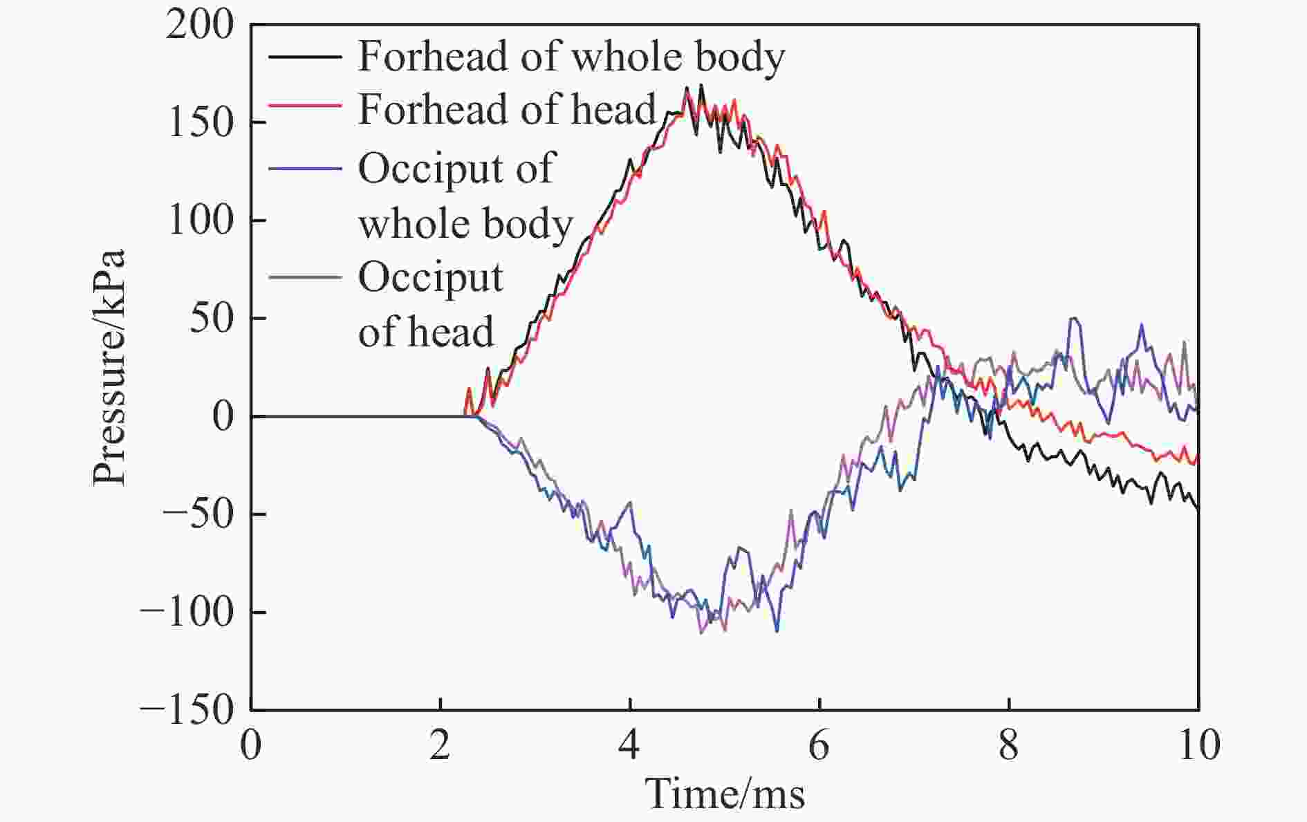
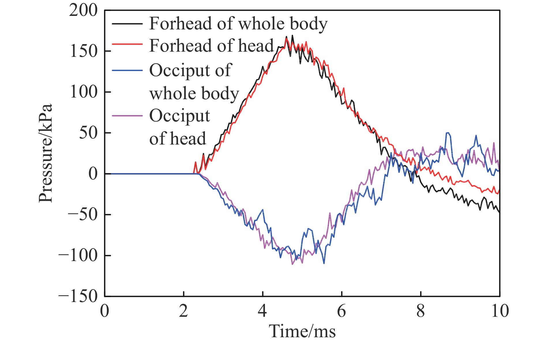
图 3 前额处与枕部处脑组织的压力曲线
Figure 3. Pressure curues of brain tissue at the forehead and occiput

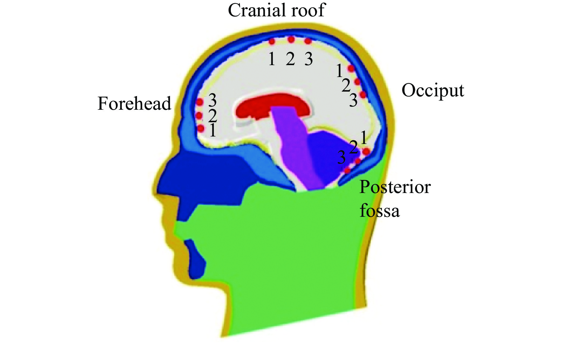
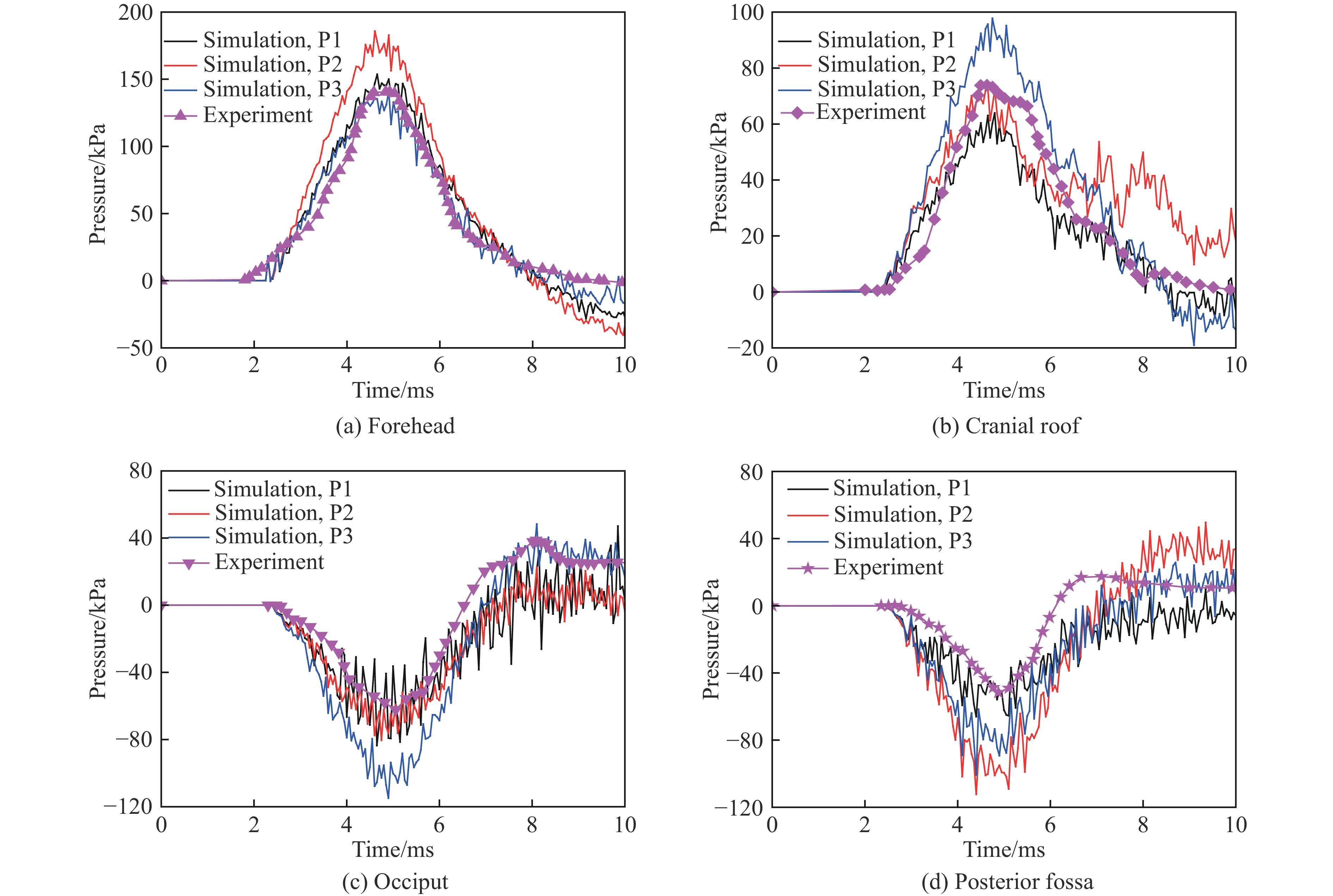
图 6 颅内压力模拟结果与实验结果的对比
Figure 6. Comparison of intracranial pressure between experiments and calculations
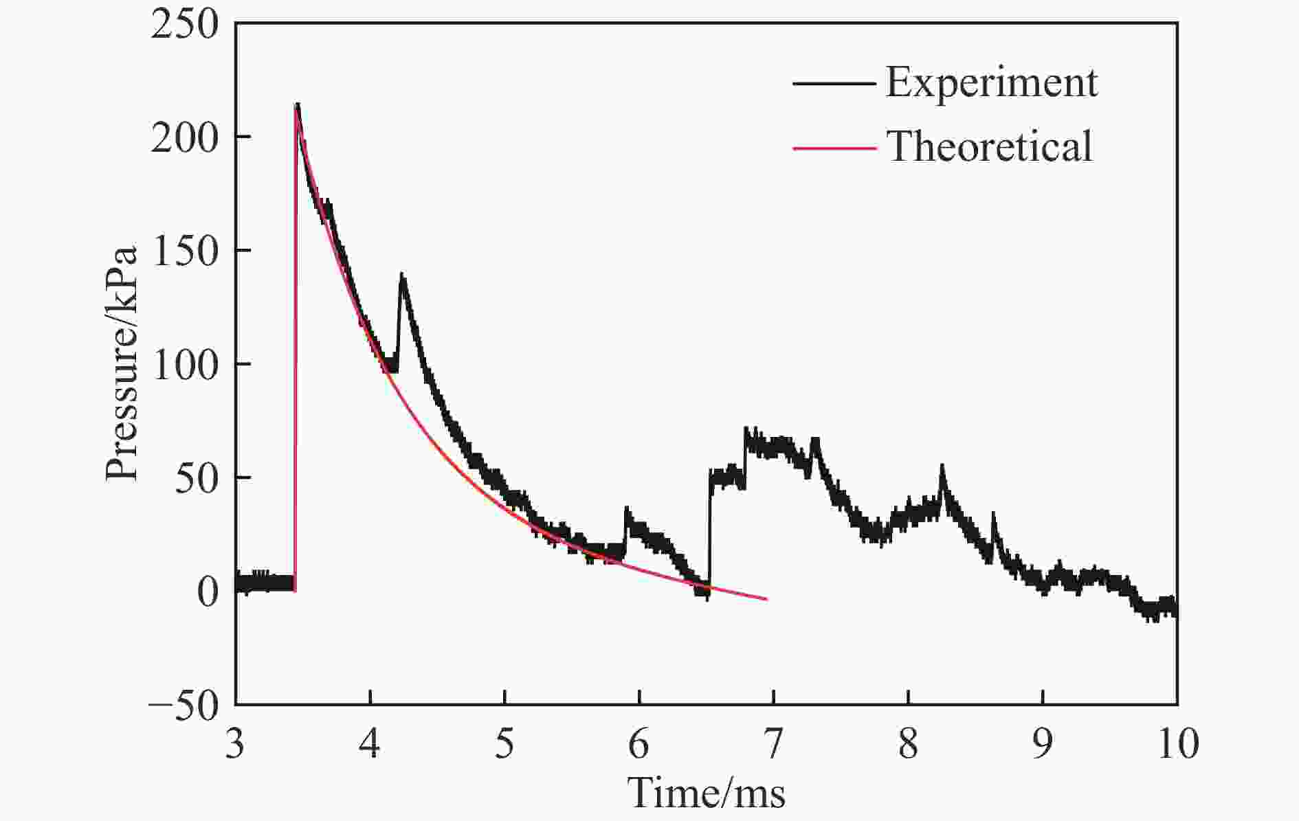
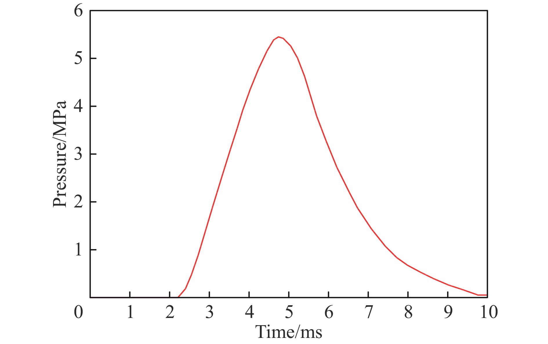
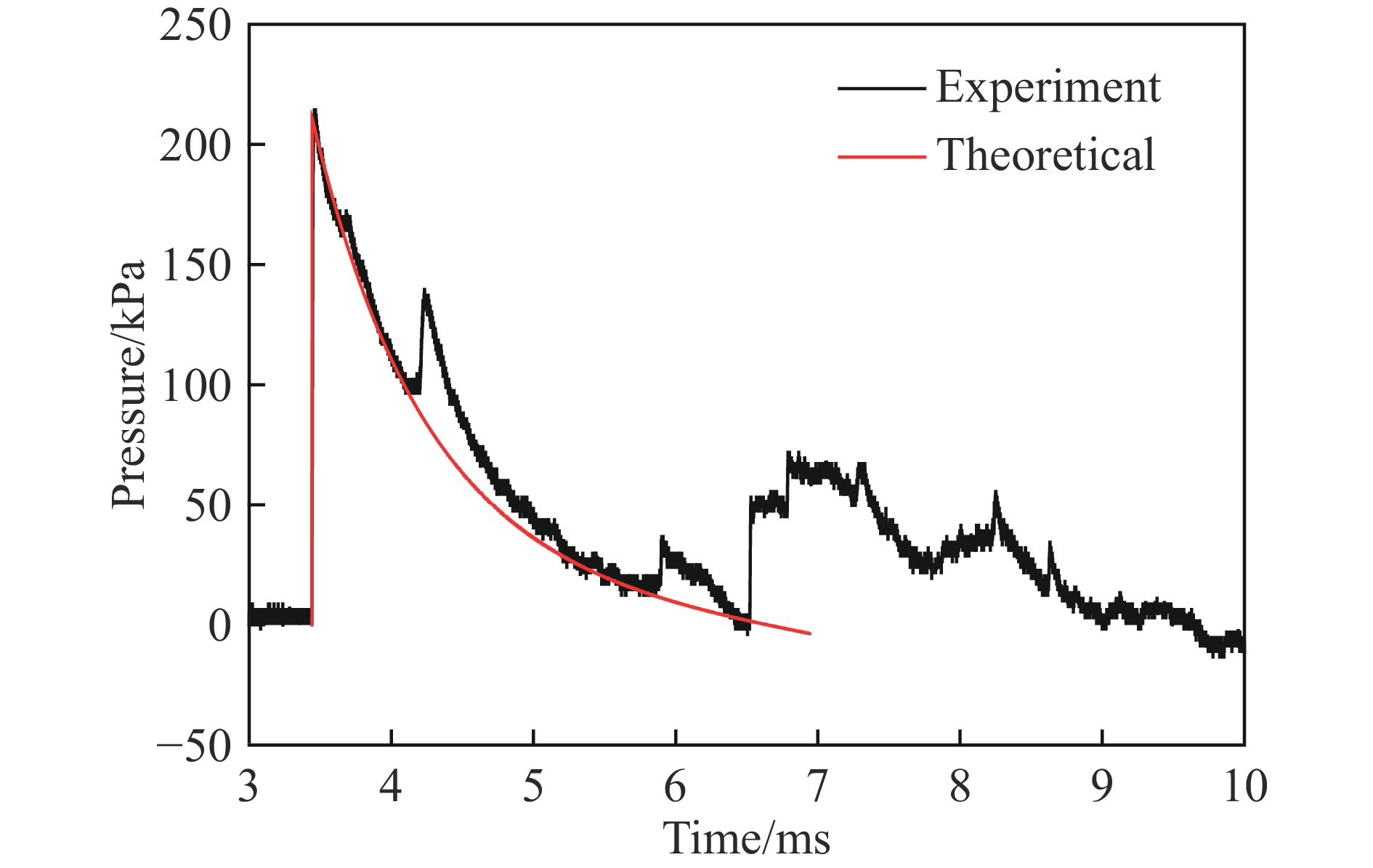
图 9 爆炸冲击波压力曲线的理论结果与实验结果对比
Figure 9. Comparison of pressure curves of explosion blast between theoretical and experimental results
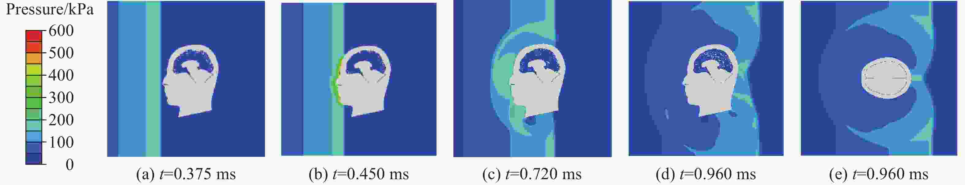
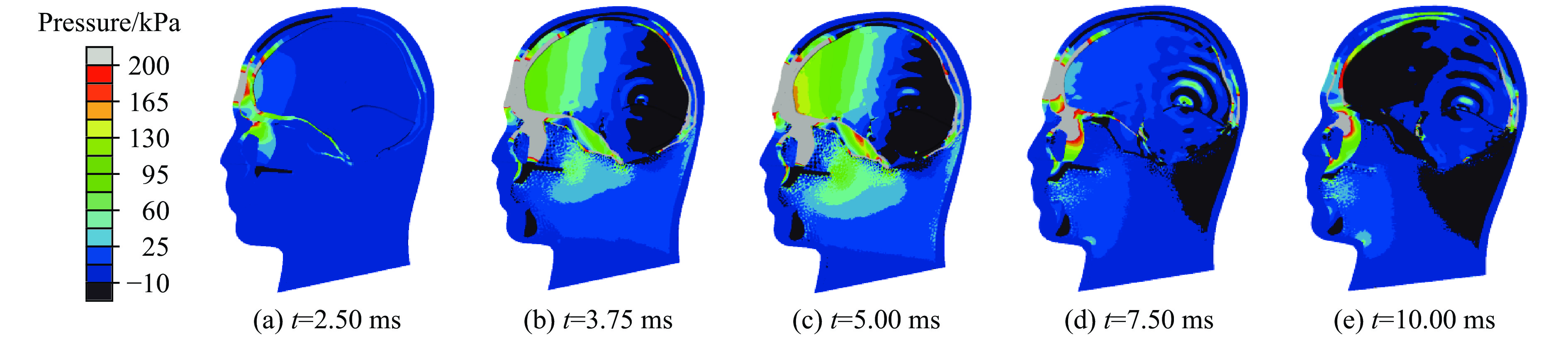
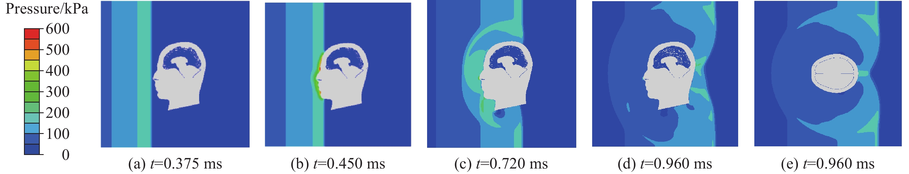
图 10 正面冲击时流场压力分布
Figure 10. Pressure distribution of flow field in blast frontal impact
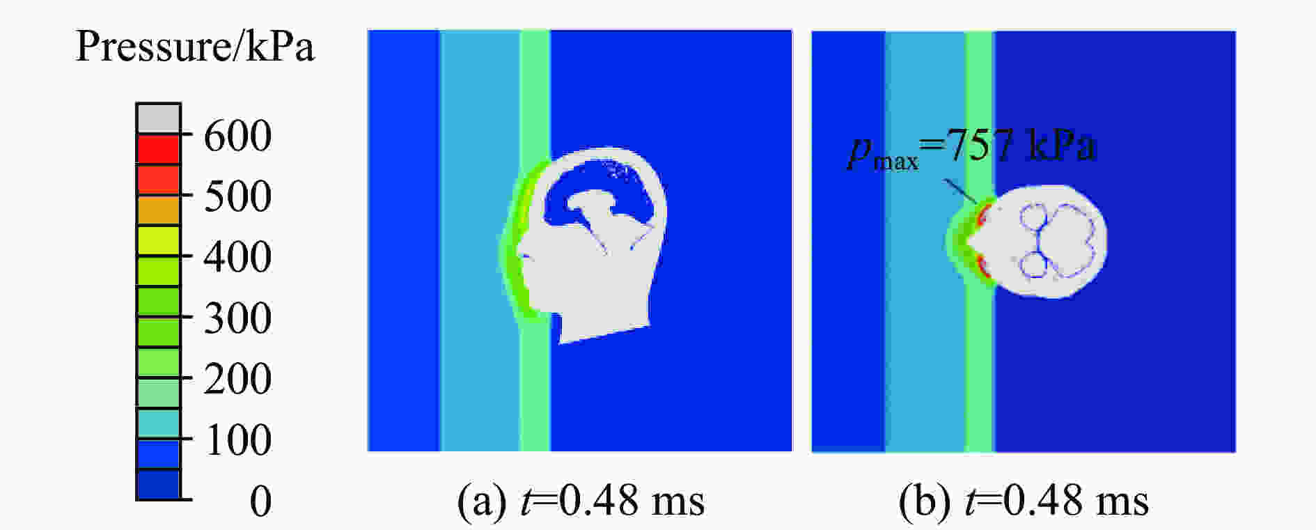
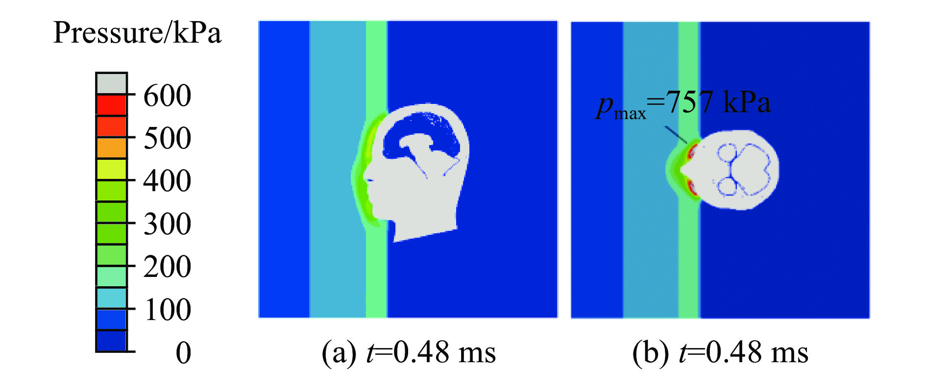
图 11 正面冲击时眼部的流场压力分布
Figure 11. Pressure distribution of flow field around the eyes in blast frontal impact

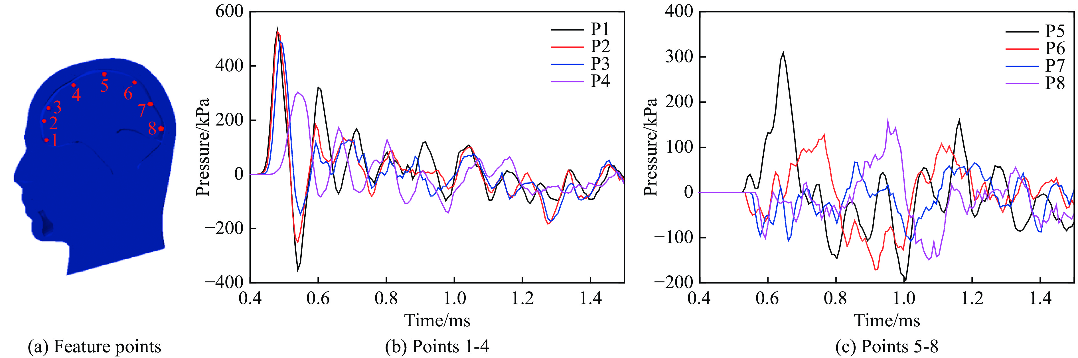
图 12 正面冲击时特征点处脑组织的压力曲线
Figure 12. Pressure curves of brain tissue at feature points in blast frontal impact
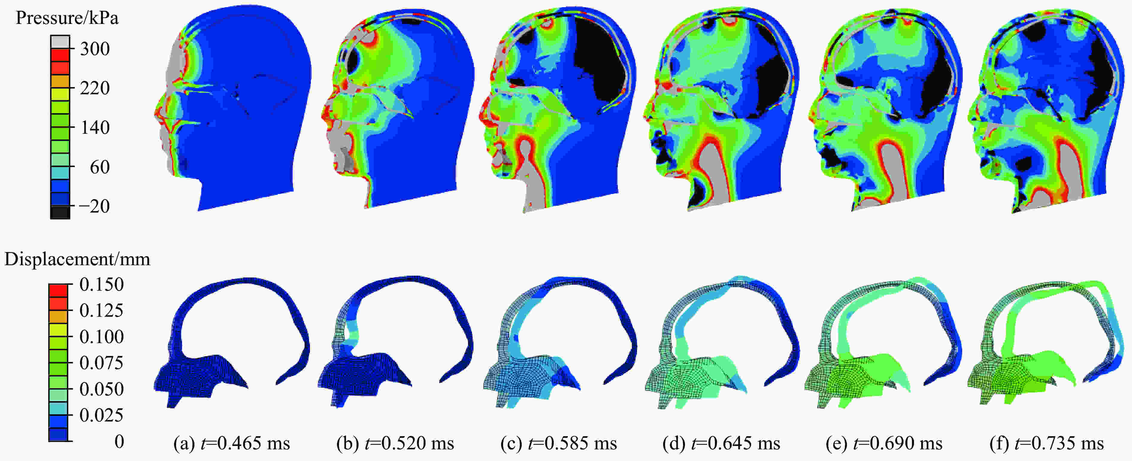
图 13 正面冲击时颅脑压力云图与颅骨变形云图
Figure 13. Nephogram of brain pressure and skull displacement in blast frontal impact
表 1 黏超弹性材料参数
Table 1. Material properties for hyper-viscoelastic model
密度/(kg·m−3) C01/Pa C10/Pa K/GPa 1 060 3 102.5 3 447.2 2.19 阶数i gi ki τi/s 1 0.528 262 0 0.008 2 0.301 899 0 0.150  下载: 导出CSV
表 2 线弹性材料参数
Table 2. Material properties for liner elastic model
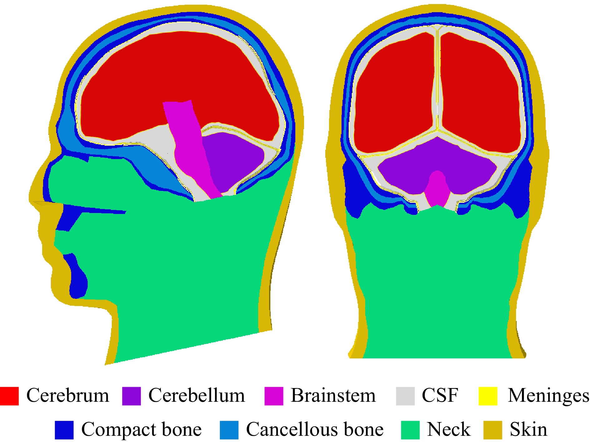
部位 密度/(kg·m−3) 弹性模量/MPa 泊松比 部位 密度/(kg·m−3) 弹性模量/MPa 泊松比 大脑镰 1 130 31.5 0.45 骨密质 2 000 15 000 0.22 硬脑膜 1 130 31.5 0.45 骨松质 1 300 1 000 0.24 小脑幕 1 130 31.5 0.45 下颌骨 1 710 5 370 0.19 蛛网膜 1 130 22.0 0.45 脖颈 1 250 354 0.30 软脑膜 1 130 11.5 0.45 皮肤 1 200 16.7 0.42
下载: 导出CSV
-
[1] TANIELIAN T, JAYCOX L H. Invisible wounds of war [R]. Santa Monica, CA: RAND Corporation, 2008. DOI: 10.1037/e527802010-001. [2] DEPALMA R G, BURRIS D G, CHAMPION H R, et al. Blast injuries [J]. New England Journal of Medicine, 2005, 352(13): 1335–1342. DOI: 10.1056/NEJMra042083. [3] MOORE D F, RADOVITZKY R A, SHUPENKO L, et al. Blast physics and central nervous system injury [J]. Future Neurology, 2008, 3(3): 243–250. DOI: 10.2217/14796708.3.3.243. [4] VERSACE J. A review of severity index [C] // Proceedings of the 15th Stapp Car Crash Conference. San Diego: Society of Automotive Engineers, 1971: 771−796. DOI: 10.4271/710881. [5] NEWMAN J A. A generalized acceleration model for brain injury threshold (GAMBIT) [C] // Proceedings of International IRCOBI Conference. 1986. [6] NEWMAN J A, SHEWCHENKO N. A proposed new biomechanical head injury assessment function: the maximum power index [R]. SAE Technical Paper, 2000. [7] CERNAK I, WANG Z G, JIANG J X, et al. Ultrastructural and functional characteristics of blast injury-induced neurotrauma [J]. The Journal of Trauma: Injury, Infection, and Critical Care, 2001, 50(4): 695–706. DOI: 10.1097/00005373-200104000-00017. [8] LIU M D, ZHANG C, LIU W B, et al. A novel rat model of blast-induced traumatic brain injury simulating different damage degree: implications for morphological, neurological, and biomarker changes [J]. Frontiers in Cellular Neuroscience, 2015, 9: 168. DOI: 10.3389/fncel.2015.00168. [9] CLOOTS R J H, VAN DOMMELEN J A W, KLEIVEN S, et al. Traumatic brain injury at multiple length scales: relating diffuse axonal injury to discrete axonal impairment [C] // IRCOBI conference. 2010. [10] CLOOTS R J H, VAN DOMMELEN J A W, GEERS M G D. A tissue-level anisotropic criterion for brain injury based on microstructural axonal deformation [J]. Journal of the Mechanical Behavior of Biomedical Materials, 2012, 5(1): 41–52. DOI: 10.1016/j.jmbbm.2011.09.012. [11] CLOOTS R J H, VAN DOMMELEN J A W, KLEIVEN S, et al. Multi-scale mechanics of traumatic brain injury: predicting axonal strains from head loads [J]. Biomechanics and Modeling in Mechanobiology, 2013, 12(1): 137–150. DOI: 10.1007/s10237-012-0387-6. [12] RADOVITZKY R, SOCRATE S, TABER K, et al. Investigations of tissue-level mechanisms of primary blast injury through modeling, simulation, neuroimaging and neuropathological studies [R]. Massachusetts Institute of Technology Cambridge, 2012. DOI: 10.21236/ada573887. [13] JEAN A, NYEIN M K, ZHENG J Q, et al. An animal-to-human scaling law for blast-induced traumatic brain injury risk assessment [J]. Proceedings of the National Academy of Sciences of the United States of America, 2014, 111(43): 15310–15315. DOI: 10.1073/pnas.1415743111. [14] GOELLER J, WARDLAW A, TREICHLER D, et al. Investigation of cavitation as a possible damage mechanism in blast-induced traumatic brain injury [J]. Journal of Neurotrauma, 2012, 29(10): 1970–1981. DOI: 10.1089/neu.2011.2224. [15] SALZAR R S, TREICHLER D, WARDLAW A, et al. Experimental investigation of cavitation as a possible damage mechanism in blast-induced traumatic brain injury in post-mortem human subject heads [J]. Journal of Neurotrauma, 2017, 34(8): 1589–1602. DOI: 10.1089/neu.2016.4600. [16] FRANCK C. Microcavitation: the key to modeling blast traumatic brain injury? [J]. Concussion, 2017, 2(3): CNC47. DOI: 10.2217/cnc-2017-0011. [17] BHATTACHARJEE Y. Shell shock revisited: solving the puzzle of blast trauma [J]. Science, 2008, 319: 406–408. DOI: 10.1126/science.319.5862.406. [18] COURTNEY A C, COURTNEY M W. A thoracic mechanism of mild traumatic brain injury due to blast pressure waves [J]. Medical Hypotheses, 2009, 72(1): 76–83. DOI: 10.1016/j.mehy.2008.08.015. [19] MOSS W C, KING M J, BLACKMAN E G. Skull flexure from blast waves: a mechanism for brain injury with implications for helmet design [J]. Physical Review Letters, 2009, 103(10): 108702. DOI: 10.1103/PhysRevLett.103.108702. [20] FELTEN D L, O’BANION M K, MAIDA M S. Netter’s atlas of neuroscience [M]. Elsevier Health Sciences, 2015: 49. [21] CHAFI M S, KARAMI G, ZIEJEWSKI M. Biomechanical assessment of brain dynamic responses due to blast pressure waves [J]. Annals of Biomedical Engineering, 2010, 38(2): 490–504. DOI: 10.1007/s10439-009-9813-z. [22] KLEIVEN S. Predictors for traumatic brain injuries evaluated through accident reconstructions [J]. Stapp Car Crash Journal, 2007, 51: 81–114. DOI: 10.12783/dtcse/wcne2017/19838. [23] GANPULE S, ALAI A, PLOUGONVEN E, et al. Mechanics of blast loading on the head models in the study of traumatic brain injury using experimental and computational approaches [J]. Biomechanics and Modeling in Mechanobiology, 2013, 12(3): 511–531. DOI: 10.1007/s10237-012-0421-8. [24] CHAFI M S, GANPULE S, GU L X, et al. Dynamic response of brain subjected to blast loadings: influence of frequency ranges [J]. International Journal of Applied Mechanics, 2011, 3(4): 803–823. DOI: 10.1142/S175882511100124X. [25] WANG C, PAHK J B, BALABAN C D, et al. Biomechanical assessment of the bridging vein rupture of blast-induced traumatic brain injury using the finite element human head model [C] // ASME 2012 International Mechanical Engineering Congress and Exposition. American Society of Mechanical Engineers, 2012: 795−805. DOI: 10.1115/imece2012-88739. [26] MOORE D F, JÉRUSALEM A, NYEIN M, et al. Computational biology:modeling of primary blast effects on the central nervous system [J]. Neuroimage, 2009, 47: T10–T20. DOI: 10.1016/j.neuroimage.2009.02.019. [27] CHEN Y, OSTOJA-STARZEWSKI M. MRI-based finite element modeling of head trauma: spherically focusing shear waves [J]. Acta Mechanica, 2010, 213(1/2): 155–167. DOI: 10.1007/s00707-009-0274-0. [28] ZOGHI-MOGHADAM M, SADEGH A M. Global/local head models to analyse cerebral blood vessel rupture leading to ASDH and SAH [J]. Computer Methods in Biomechanics and Biomedical Engineering, 2009, 12(1): 1–12. DOI: 10.1080/10255840802020420. [29] NAHUM A M, SMITH R, WARD C C. Intracranial pressure dynamics during head impact [C] // Proceedings of 21st Stapp Car Crash Conference. Pennsylvania: Society of Automotive Engineers, 1977: 339−366. DOI: 10.4271/770922. [30] BENEDICT J V, HARRIS E H, VON ROSENBERG D U. An analytical investigation of the cavitation hypothesis of brain damage [J]. Journal of Basic Engineering, 1970, 92(3): 597–603. DOI: 10.1115/1.3425083. [31] WARD C, CHAN M, NAHUM A. Intracranial pressure: a brain injury criterion [R]. SAE Technical Paper, 1980. DOI: 10.4271/801304. -


计量
- 文章访问数: 5994
- HTML全文浏览量: 2325
- PDF下载量: 192
- 被引次数: 0