



Dynamic mechanical properties and fracture mechanism of cortical bone under impact loading
-
摘要: 借助万能试验机和分离式霍普金森压杆装置对鸡股骨皮质骨开展了准静态(0.001 s−1)与动态(350和700 s−1)单轴压缩实验,结合数字图像相关技术获取全场应变分布,并利用扫描电镜观察断裂微观形貌,分析了应变率对断裂行为的影响。实验结果表明,皮质骨的破坏应力随应变率升高成非单调变化,在0.001、350和700 s−1下平均值分别为99.6、195.5和171.3 MPa,表现出“先增后降”的趋势,断裂应变也有类似趋势。在准静态加载下,试样宏观裂纹路径与最大切应变区域一致,断裂面呈现层状剥离和大量碎屑等典型剪切破坏特征,表明准静态下皮质骨的断裂机制由剪切应变主导;而在动态加载下,宏观裂纹更倾向于与最大拉应变区域吻合,断裂面较为光滑,呈现典型拉伸断裂形貌,表明动态条件下拉应变起主导作用。这说明皮质骨断裂机制随应变率的升高,由剪切主导向拉伸主导的转变。Abstract: An uniaxial compression tests were conducted on chicken femoral cortical bone under quasi-static (0.001 s−1) and dynamic (350 and 700 s−1) conditions by using a universal testing machine and a split Hopkinson pressure bar (SHPB). Chicken femoral cortical bone specimens were obtained via cutting and grinding, with high-contrast speckle patterns prepared on them. Images of cortical bone were recorded during the experiment using two cameras, with an acquisition frequency of 5 Hz for the quasi-static test and 150 kHz for the dynamic test. Full-field strain distributions were obtained using digital image correlation (DIC), and fracture micro-morphology was observed via scanning electron microscopy (SEM) to analyze the effect of strain rate on fracture behavior. The experimental results show that the failure stress of cortical bone exhibits a non-monotonic trend with increasing strain rate, with average values of 99.6, 195.5 and 171.3 MPa at 0.001, 350, and 700 s−1, respectively, demonstrating an “increase-then-decrease” tendency. A similar trend was observed for fracture strain. Under quasi-static loading, the macroscopic crack propagation path aligns with the maximum shear strain region, and the fracture surface exhibits typical shear failure characteristics (layered delamination and abundant debris), indicating shear strain as the primary fracture driver. In contrast, under dynamic loading, the macroscopic crack correlates more closely with the maximum tensile strain region, and the fracture surface appears smooth, characteristic of tensile fracture morphology, confirming tensile strain as the dominant mechanism. Strain rate exerts a significant influence on the fracture mechanism by regulating the fracture-inducing factor, which may account for one of the key reasons for the variation in fracture stress of cortical bone under dynamic conditions. This study reveals, from a macro-micro correlated perspective, the shift in fracture mechanisms of cortical bone from shear dominance to tensile dominance with increasing strain rate, which provides experimental evidence for understanding the fracture mechanism of bone under impact loading and a reference for the design of bone biomimetic materials and the development of orthopedic protective equipment.
-
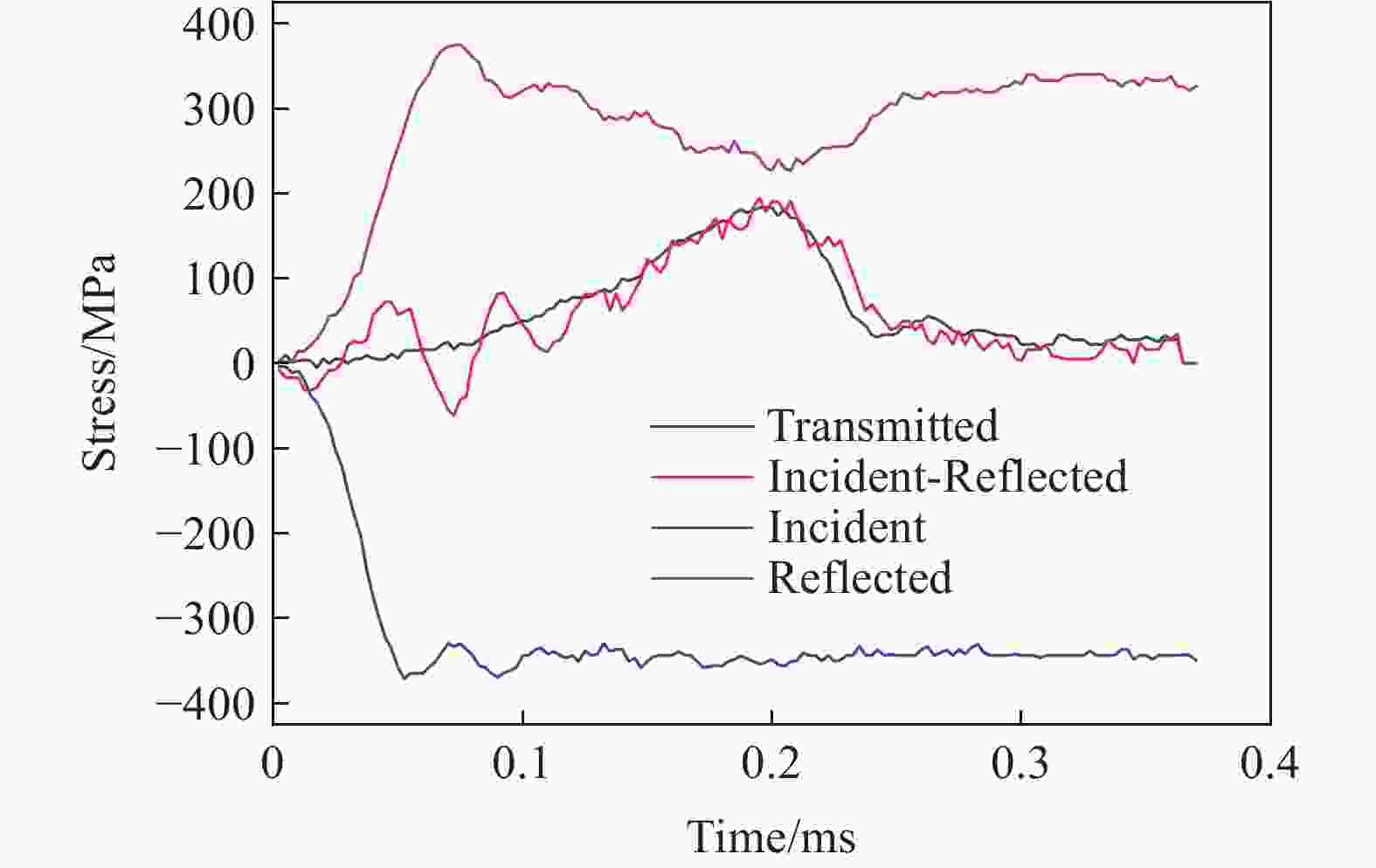
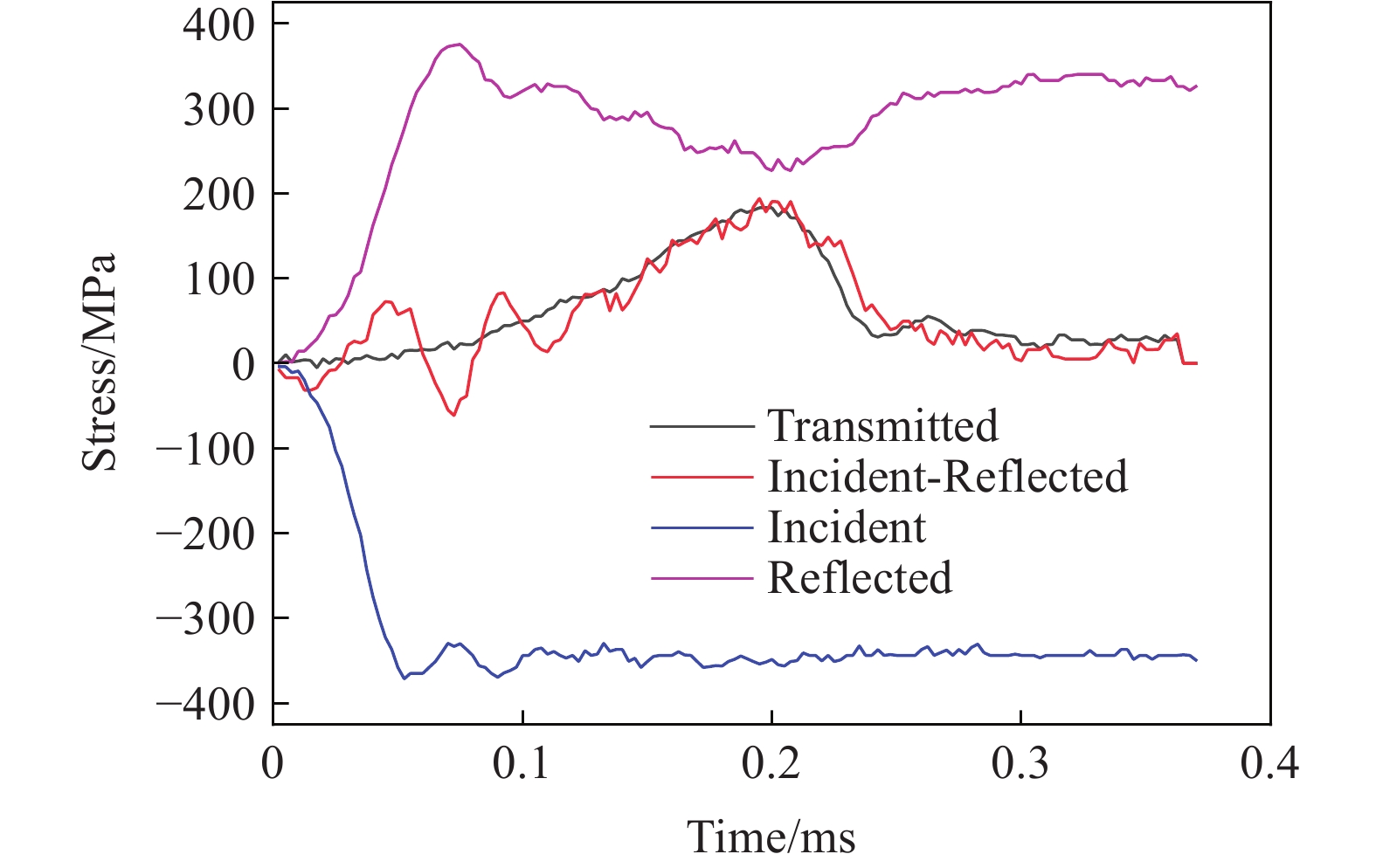
图 3 入射波、反射波、透射波(T)及入射波与反射波之差(I-R)
Figure 3. Incident wave, reflected wave, transmitted wave (T), and the difference between incident and reflected waves (I-R)
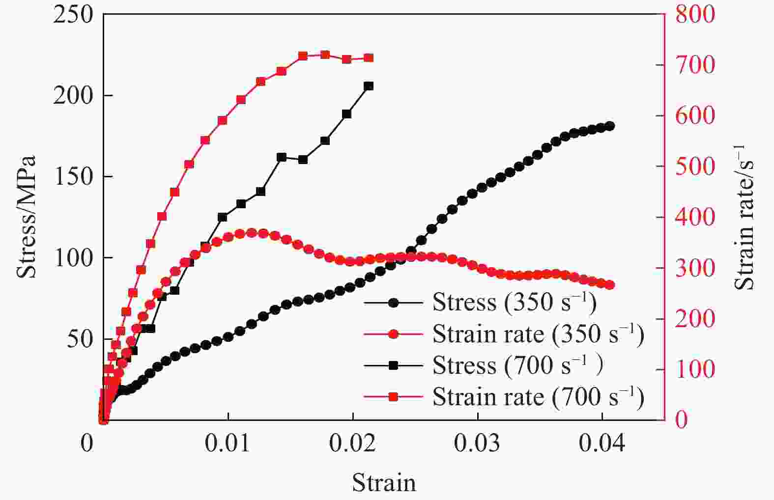
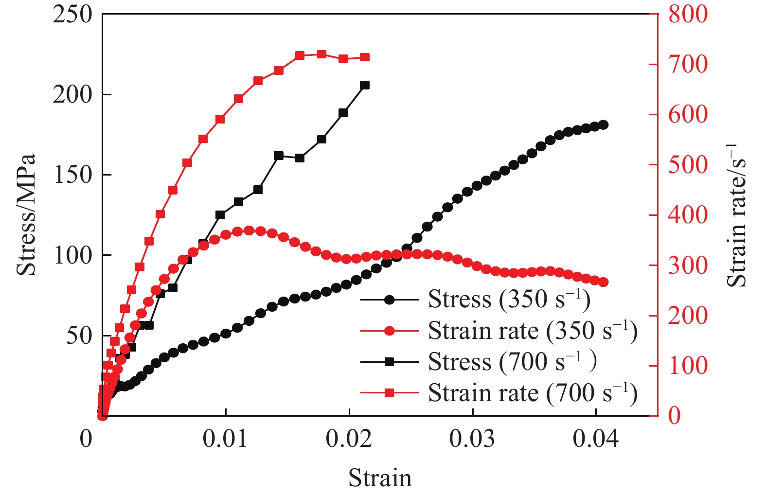
图 4 动态实验中的典型应力-应变及应变率-应变曲线图
Figure 4. Typical stress-strain and strain rate-strain curves in dynamic experiments
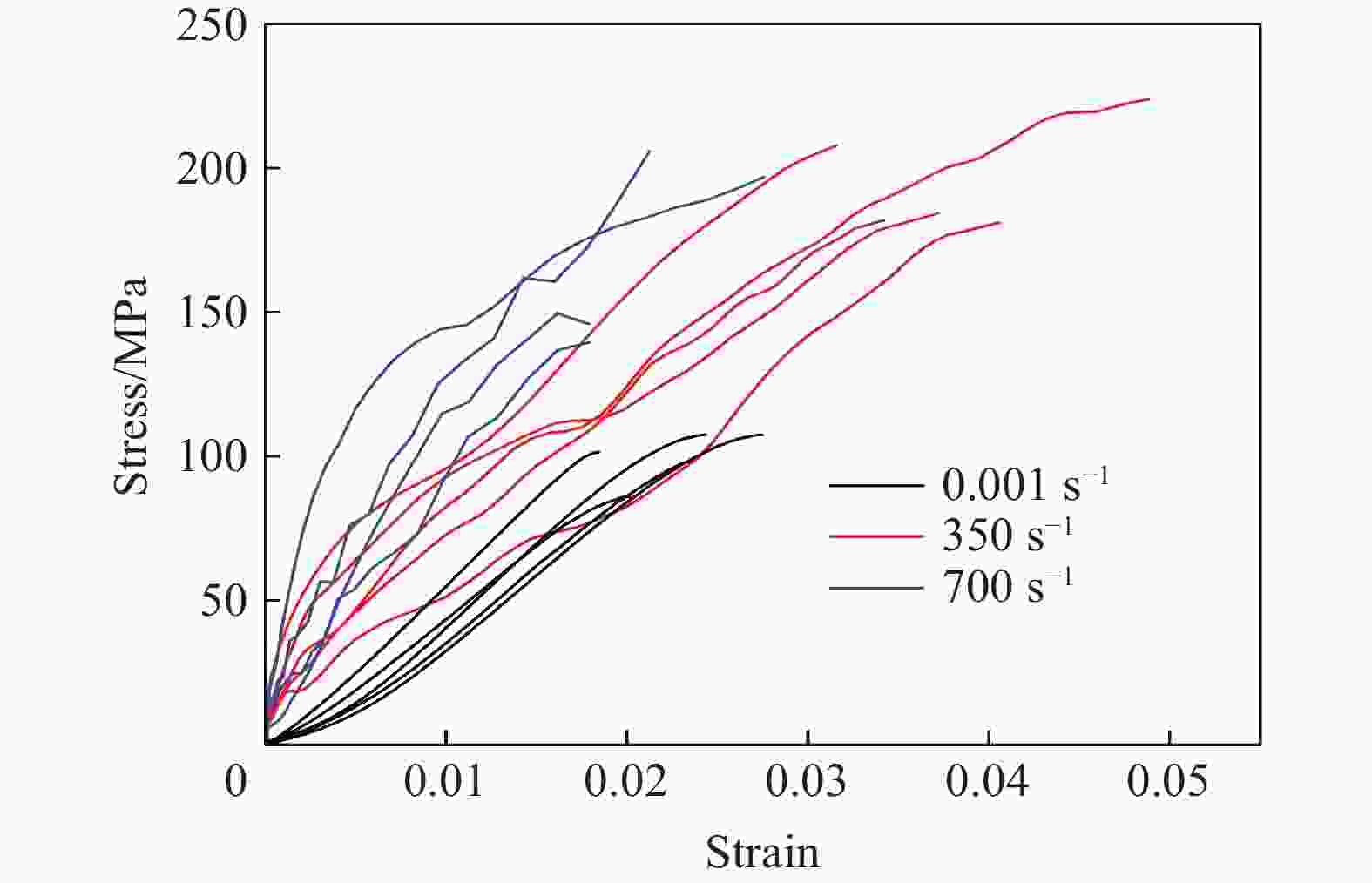
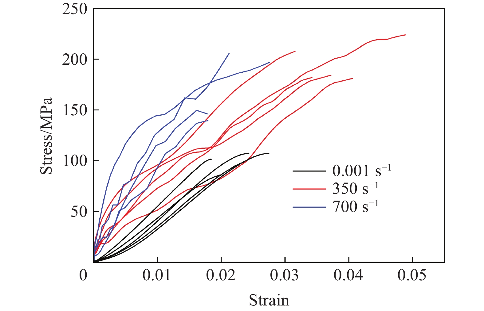
图 5 三种应变率条件下的应力-应变曲线对比
Figure 5. Comparison of stress-strain curves under three different strain rate conditions
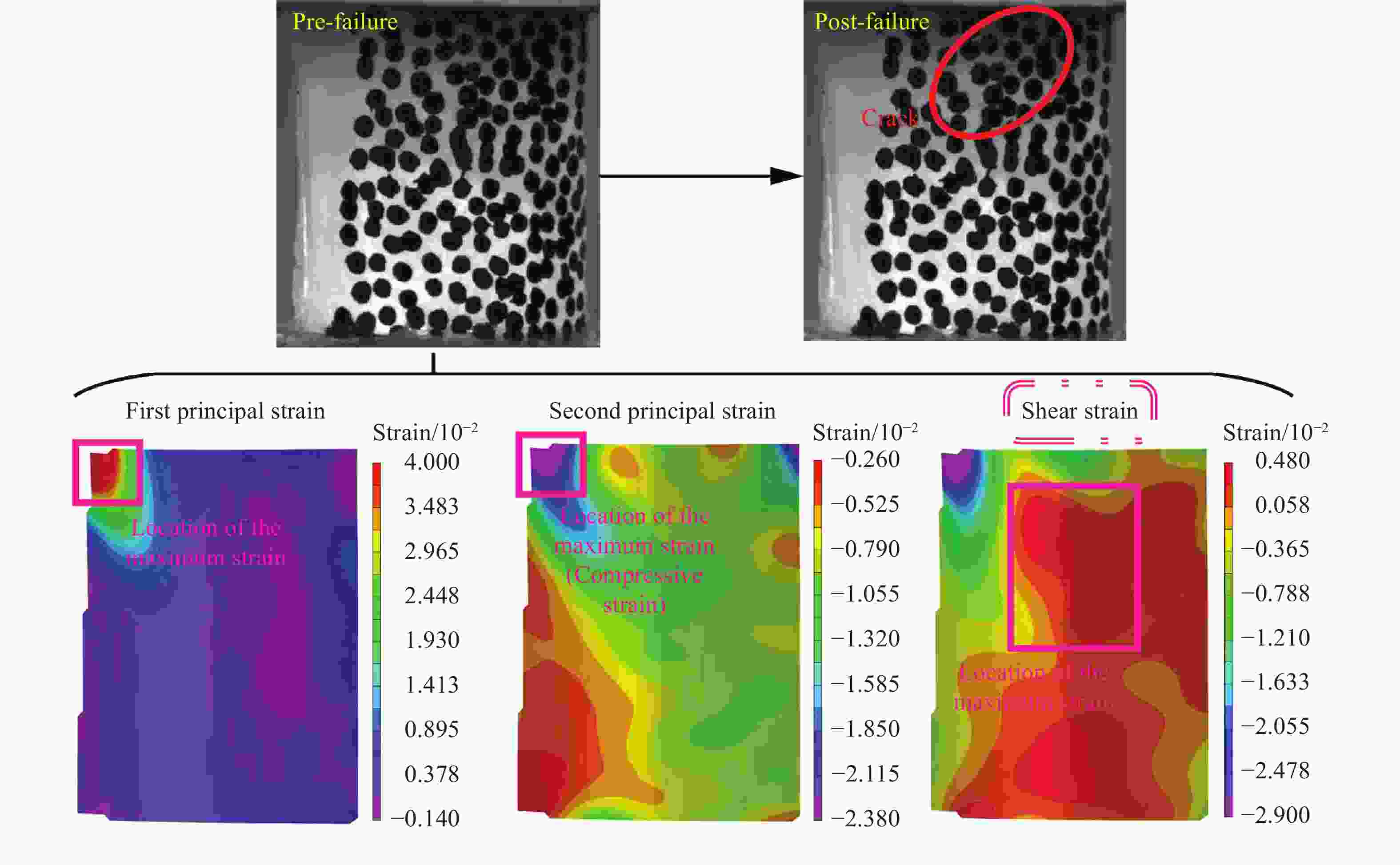
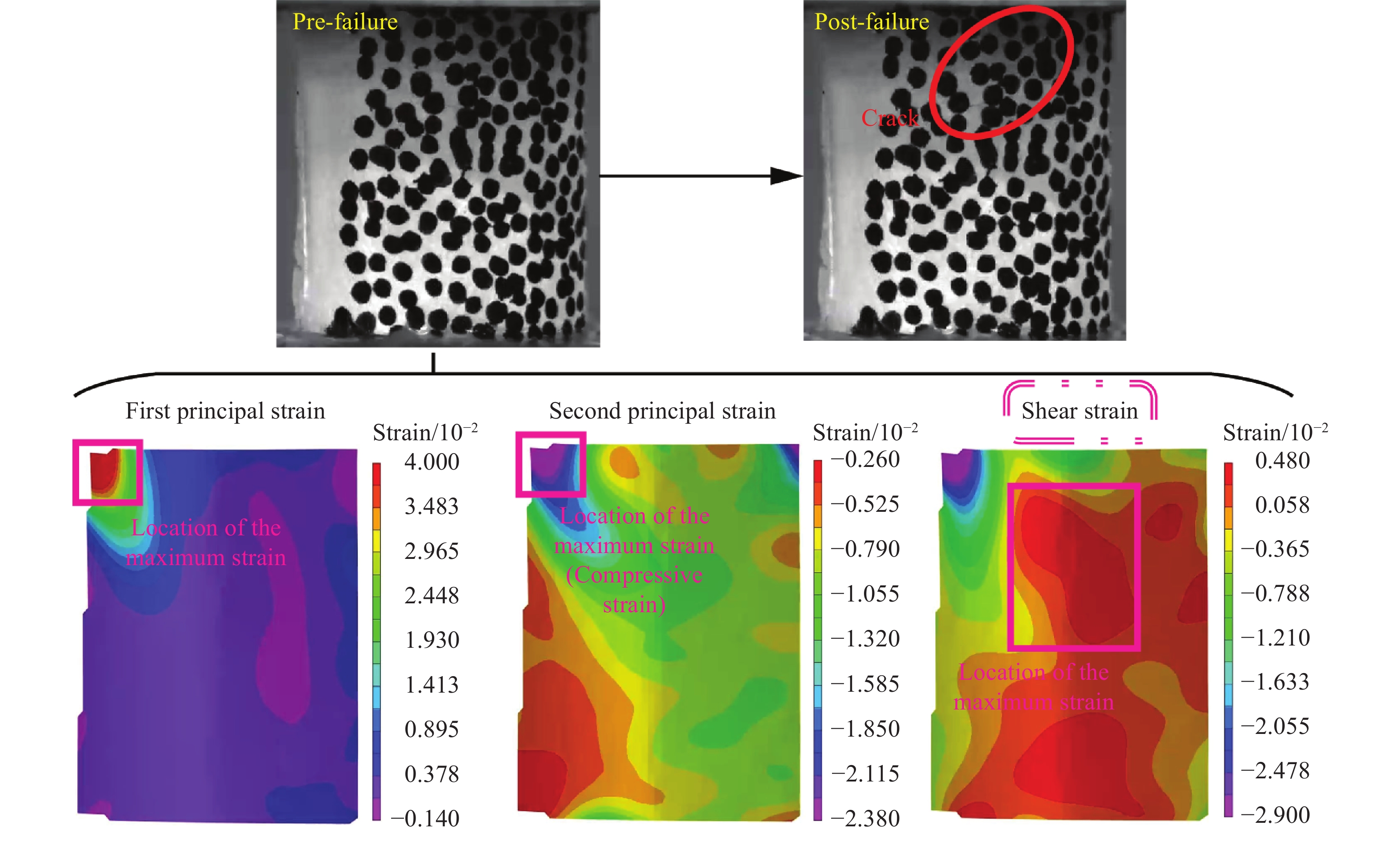
图 6 应变率为0.001 s−1时皮质骨试样破坏前一刻的应变云图及裂纹萌生位置
Figure 6. Strain field and the location of crack nucleation of the cortical bone specimen under quasi-static loading (0.001 s−1)
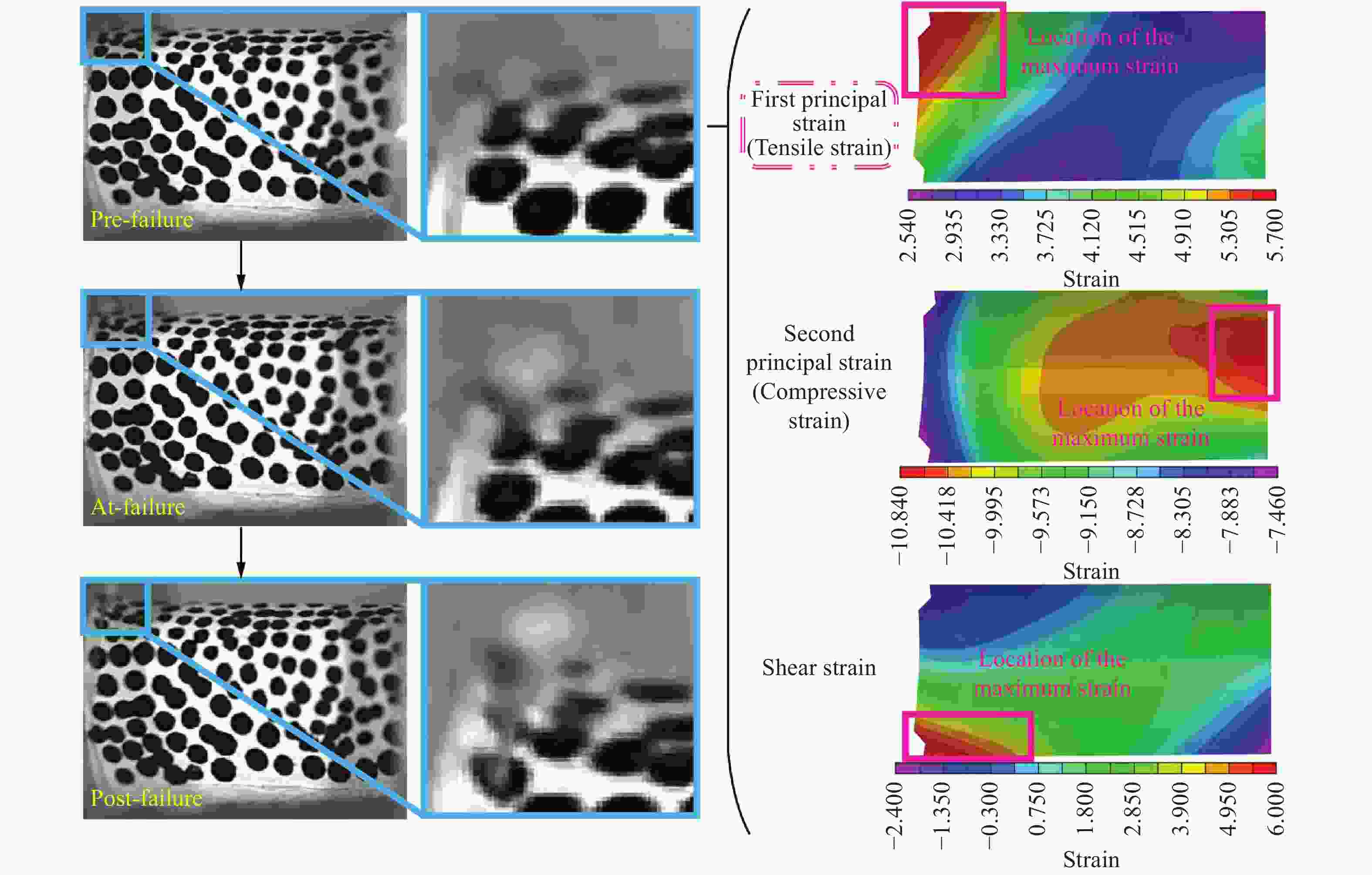
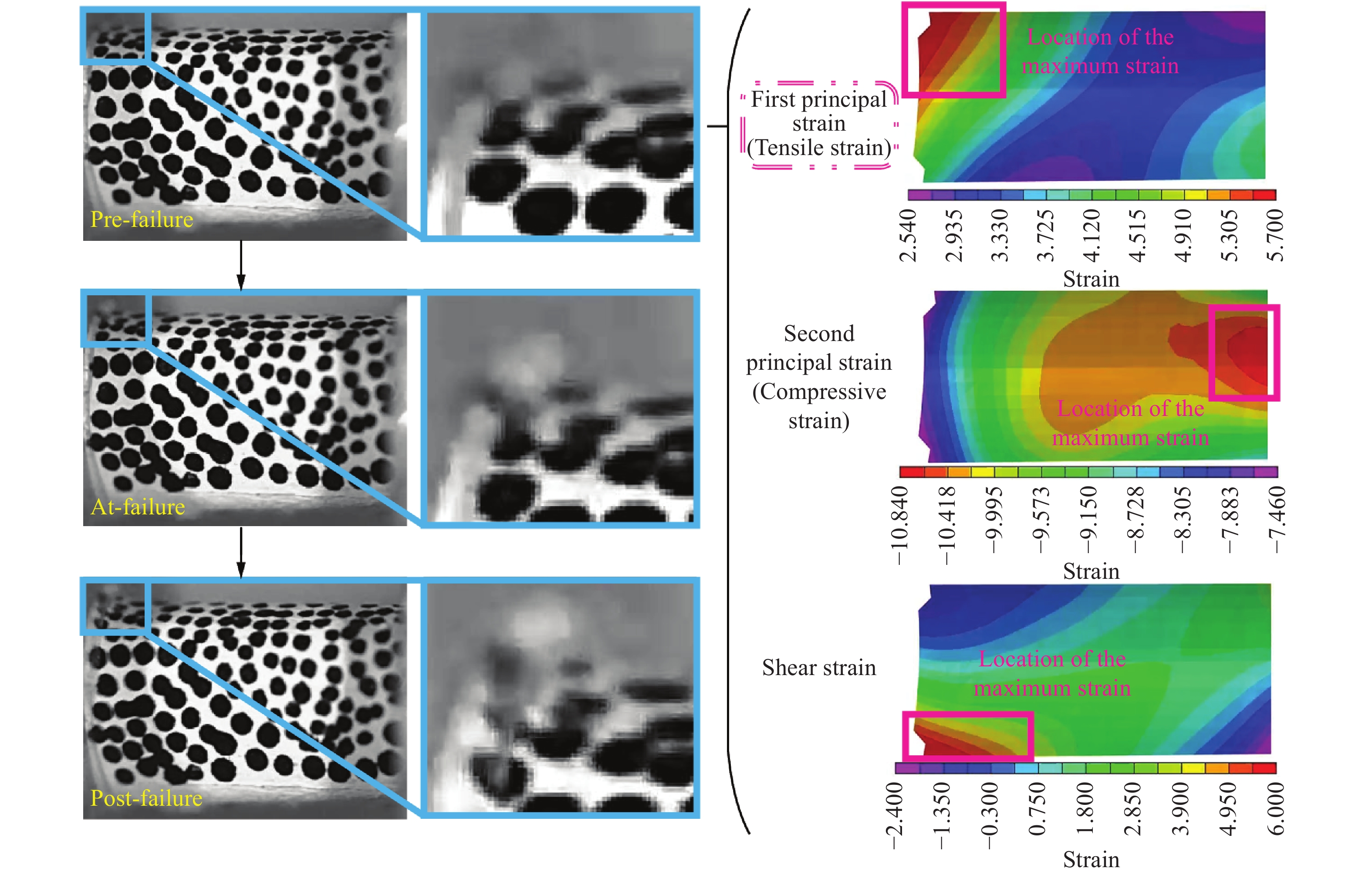
图 7 应变率为350 s−1时皮质骨试样破坏前一刻的应变云图及裂纹萌生位置
Figure 7. Strain field and the location of crack nucleation of the cortical bone specimen under the loading with the strain rate of 350 s−1
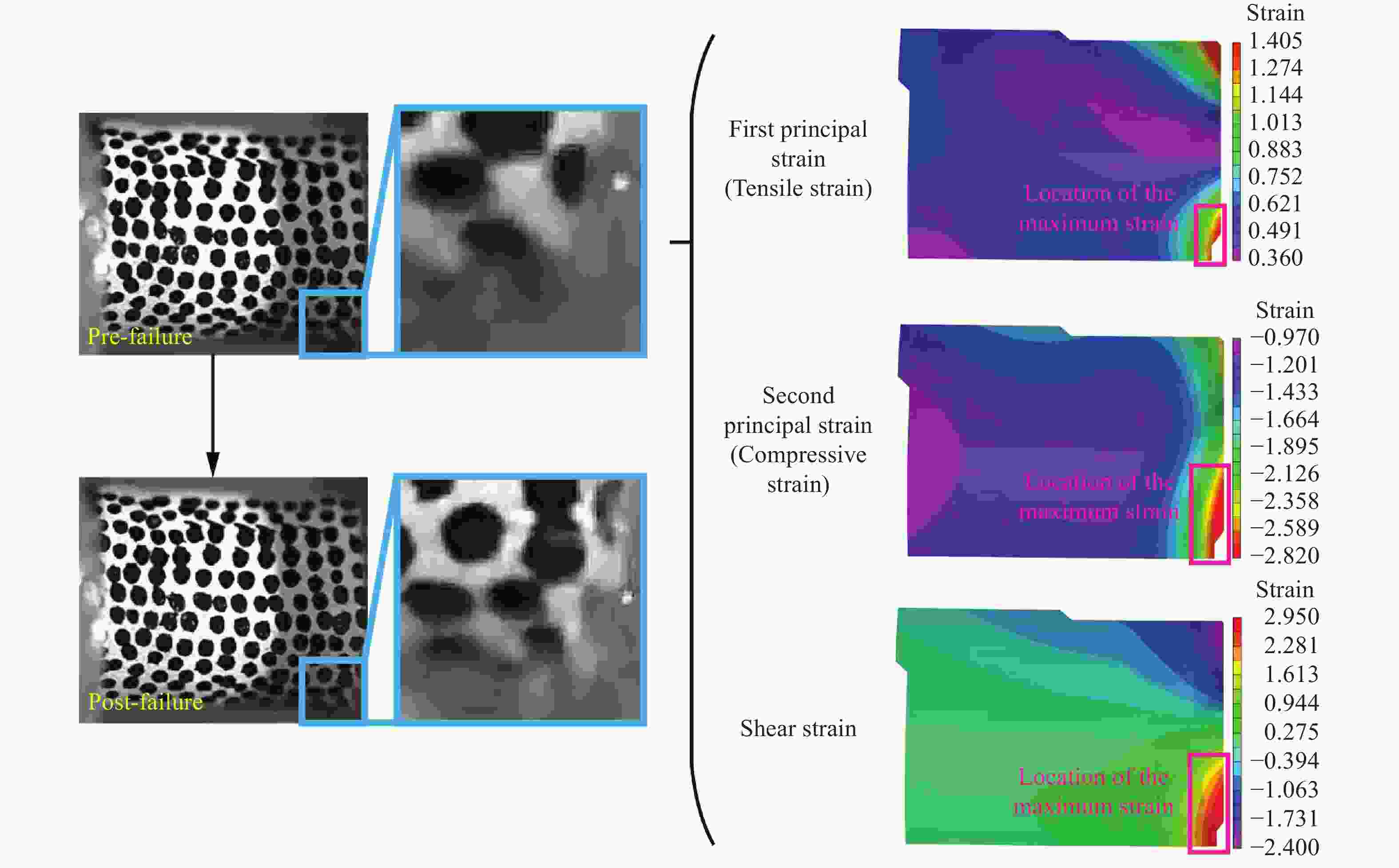
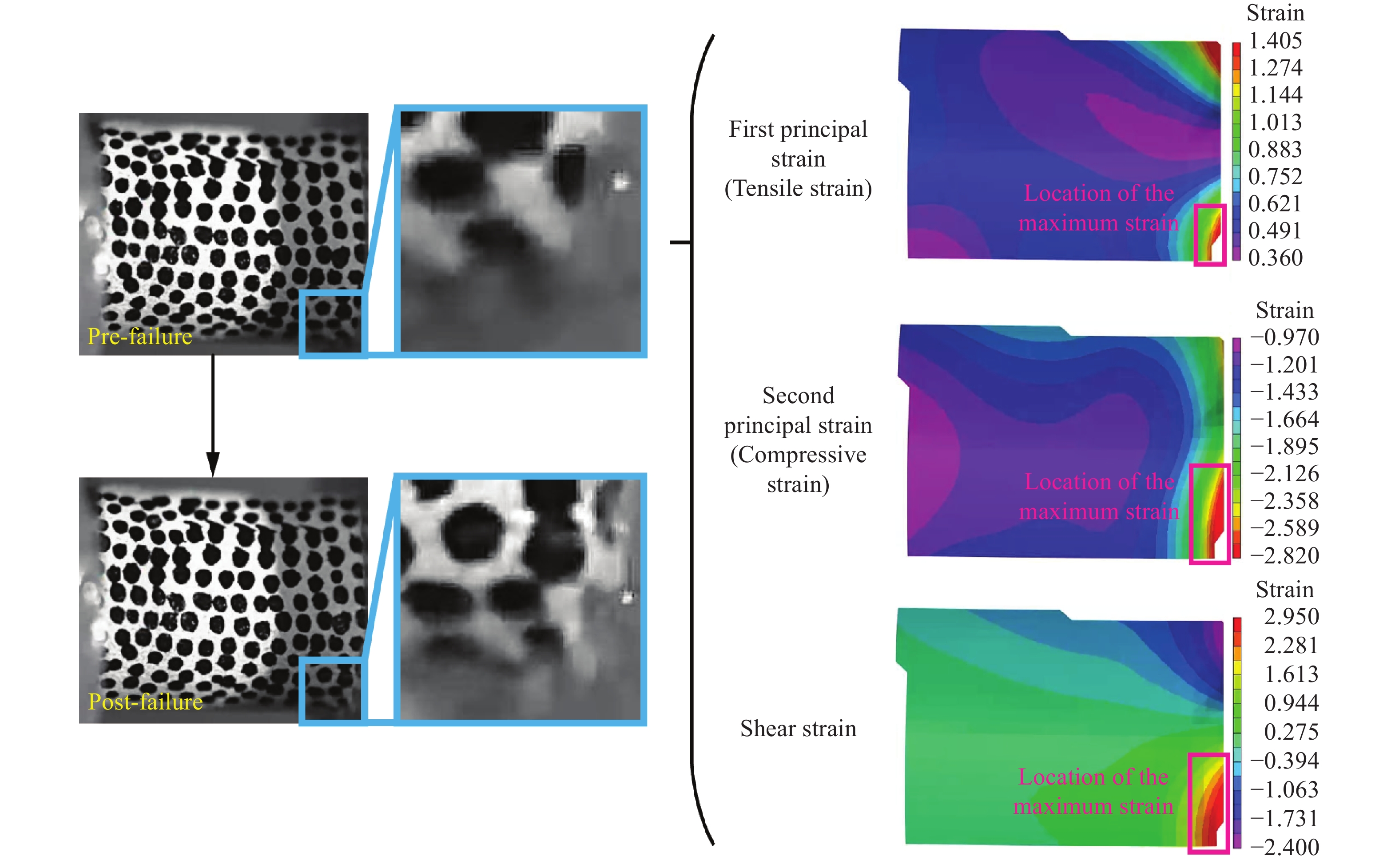
图 8 应变率为700 s−1时皮质骨试样破坏前一刻的应变云图及裂纹萌生位置
Figure 8. Strain field and the location of crack nucleation of the cortical bone specimen under the loading with the strain rate of 700 s−1
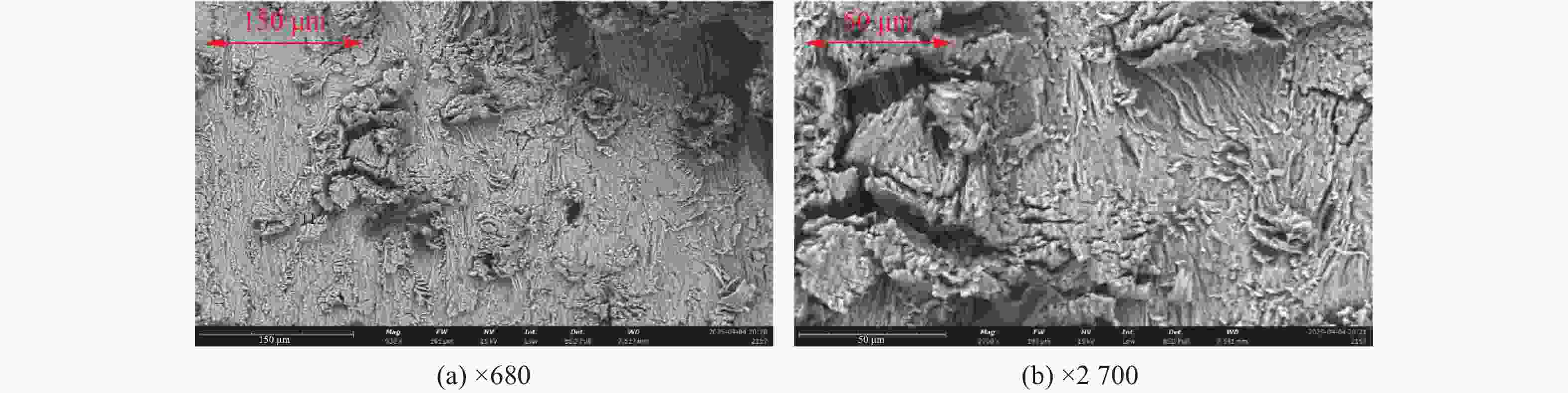
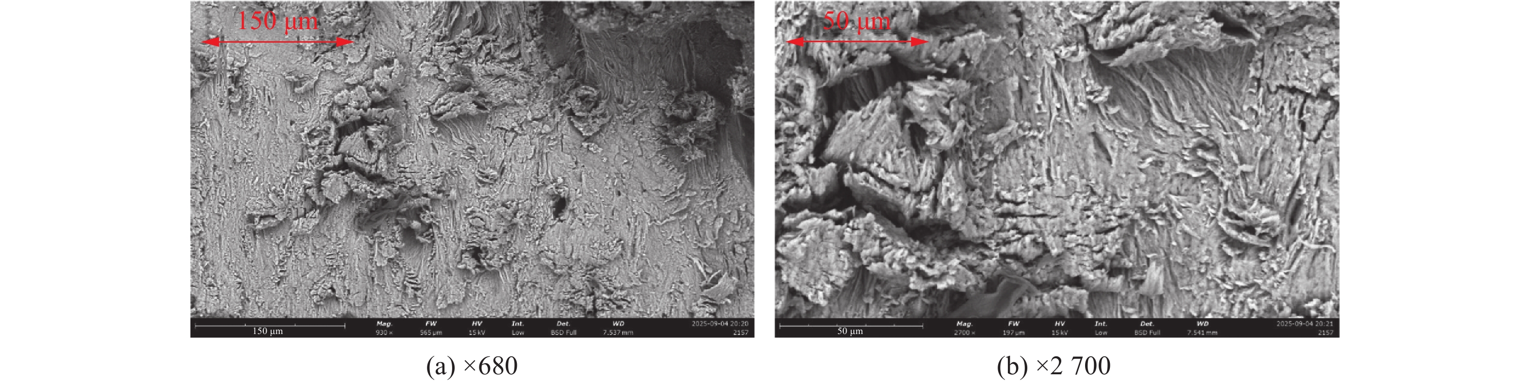
图 9 准静态条件下断裂面的SEM图像
Figure 9. SEM images of the fracture surface under quasi-static conditions
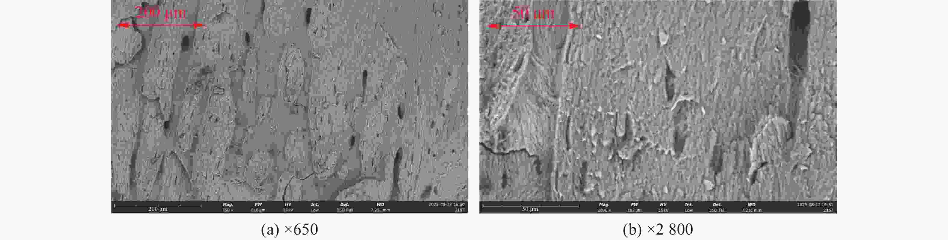
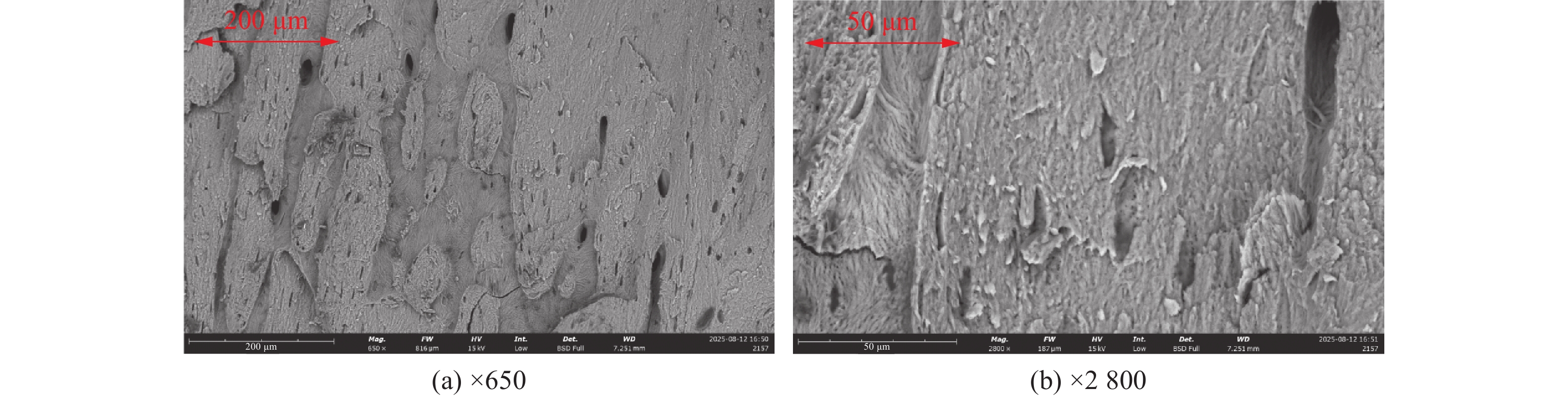
图 10 350s−1应变率条件下断裂面的扫描电镜SEM图像
Figure 10. SEM images of the fracture surface under the loading with the strain rate of 350 s−1
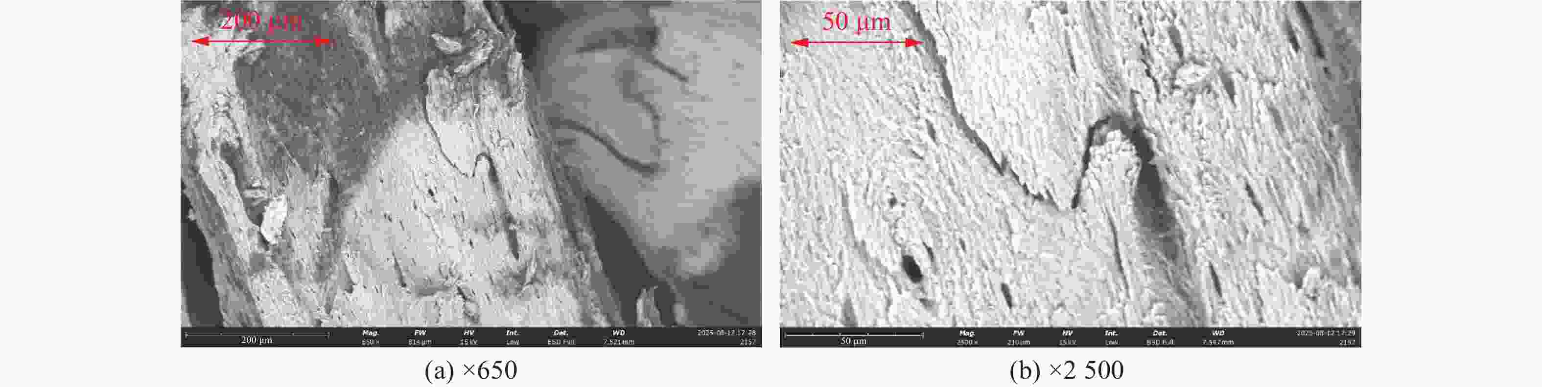
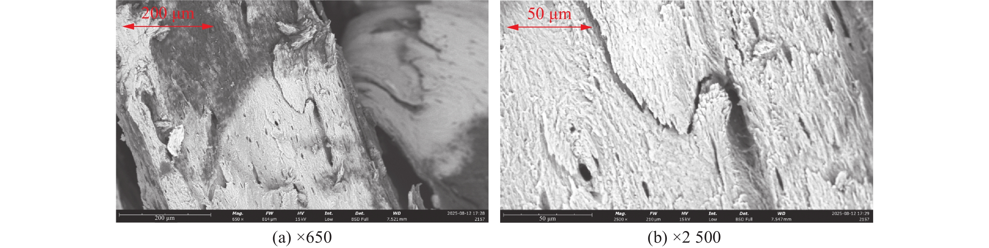
图 11 700 s−1应变率条件下断裂面的扫描电镜SEM图像
Figure 11. SEM images of the fracture surface under the loading with the strain rate of 700 s−1
表 1 三种应变率条件下对应的平均破坏应力与平均弹性模量
Table 1. Average failure stress and elastic modulus under the three strain rates
加载工况 平均破坏应力/MPa 平均弹性模量/GPa 准静态 99.6 (n=5, μ=8.1) 5.255 (n=5, μ=0.402) 350 s−1 195.5 (n=5, μ=16.9) \ 700 s−1 171.3 (n=4, μ=26.4) \  下载: 导出CSV
-
[1] BLACK J D J, TADROS B J. Bone structure: from cortical to calcium [J]. Orthopaedics and Trauma, 2020, 34(3): 113–119. DOI: 10.1016/j.mporth.2020.03.002. [2] 许灿, 李明清, 王成功, 等. 皮质骨的多级结构与韧化机制研究进展 [J]. 医用生物力学, 2018, 33(3): 273–279. DOI: 10.16156/j.1004-7220.2018.03.014.XU C, LI M Q, WANG C G, et al. Research progress on hierarchical structure of cortical bone and its toughening mechanisms [J]. Journal of Medical Biomechanics, 2018, 33(3): 273–279. DOI: 10.16156/j.1004-7220.2018.03.014. [3] DAPAAH D, MARTEL D R, LAING A C, et al. The impact of fall-related loading rate on the formation of micro-damage in human cortical bone fracture [J]. Journal of Biomechanics, 2022, 142: 111254. DOI: 10.1016/j.jbiomech.2022.111254. [4] GAUTHIER R, FOLLET H, LANGER M, et al. Strain rate influence on human cortical bone toughness: a comparative study of four paired anatomical sites [J]. Journal of the Mechanical Behavior of Biomedical Materials, 2017, 71: 223–230. DOI: 10.1016/j.jmbbm.2017.03.015. [5] QIU J L, LIAO Z K, XIANG H Y, et al. Effects of different preservation on the mechanical properties of cortical bone under quasi-static and dynamic compression [J]. Frontiers in Bioengineering and Biotechnology, 2023, 11: 1082254. DOI: 10.3389/fbioe.2023.1082254. [6] 徐诚意, 刘坤, 康宝, 等. 不同应变率下皮质骨压缩力学实验与本构模型 [J]. 爆炸与冲击, 2025, 45(7): 071426. DOI: 10.11883/bzycj-2024-0513.XU C Y, LIU K, KANG B, et al. Study on compressive mechanical tests and constitutive models of cortical bone under different strain rates [J]. Explosion and Shock Waves, 2025, 45(7): 071426. DOI: 10.11883/bzycj-2024-0513. [7] KUMAR A, GHOSH R. A review on experimental and numerical investigations of cortical bone fracture [J]. Proceedings of the Institution of Mechanical Engineers, Part H: Journal of Engineering in Medicine, 2022, 236(3): 297–319. DOI: 10.1177/09544119211070347. [8] ALLAHYARI P, SILANI M, YAGHOUBI V, et al. On the fracture behavior of cortical bone microstructure: the effects of morphology and material characteristics of bone structural components [J]. Journal of the Mechanical Behavior of Biomedical Materials, 2023, 137: 105530. DOI: 10.1016/j.jmbbm.2022.105530. [9] KUMAR A, PATHAK H, GHOSH R. Cortical bone fracture analysis including the combined influence of osteon orientations, applied load and crack lengths: a numerical investigation [J]. Proceedings of the Institution of Mechanical Engineers, Part H: Journal of Engineering in Medicine, 2024, 238(11/12): 1091–1102. DOI: 10.1177/09544119241299081. [10] MAGHAMI E, JOSEPHSON T O, MOORE J P, et al. Fracture behavior of human cortical bone: role of advanced glycation end-products and microstructural features [J]. Journal of Biomechanics, 2021, 125: 110600. DOI: 10.1016/j.jbiomech.2021.110600. [11] GUSTAFSSON A, KHAYYERI H, WALLIN M, et al. An interface damage model that captures crack propagation at the microscale in cortical bone using XFEM [J]. Journal of the Mechanical Behavior of Biomedical Materials, 2019, 90: 556–565. DOI: 10.1016/j.jmbbm.2018.09.045. [12] PÉREZ-CANO F D, PARRA-CABRERA G, JIMÉNEZ-DELGADO J J. An approach to microscopic cortical bone fracture simulation: enhancing clinical replication [J]. International Journal of Computational Intelligence Systems, 2024, 17(1): 102. DOI: 10.1007/s44196-024-00486-9. [13] GUSTAFSSON A, GALTERI G, BARAKAT A, et al. Characterization of damage mechanisms in cortical bone: quantification of fracture resistance, critical strains, and crack tortuosity [J]. Journal of the Mechanical Behavior of Biomedical Materials, 2024, 160: 106721. DOI: 10.1016/j.jmbbm.2024.106721. [14] SUN X D, WU W D, ZHANG R B, et al. Mechanical response and in-situ deformation mechanism of cortical bone materials under combined compression and torsion loads [J]. PLoS One, 2022, 17(7): e0271301. DOI: 10.1371/journal.pone.0271301. [15] ZHAI X D, NIE Y Z, GAO J L, et al. The effect of loading direction on the fracture behaviors of cortical bone at a dynamic loading rate [J]. Journal of the Mechanics and Physics of Solids, 2020, 142: 104015. DOI: 10.1016/j.jmps.2020.104015. [16] 安兵兵, 李凯, 张东升. 皮质骨断裂力学行为的实验研究 [J]. 力学学报, 2010, 42(6): 1164–1171. DOI: 10.6052/0459-1879-2010-6-lxxb2009-604.AN B B, LI K, ZHANG D S. An experimental approach on fracture behavior of cortical bones [J]. Chinese Journal of Theoretical and Applied Mechanics, 2010, 42(6): 1164–1171. DOI: 10.6052/0459-1879-2010-6-lxxb2009-604. [17] SHUNMUGASAMY V C, GUPTA N, COELHO P G. High strain rate response of rabbit femur bones [J]. Journal of Biomechanics, 2010, 43(15): 3044–3050. DOI: 10.1016/j.jbiomech.2010.06.034. [18] ADHARAPURAPU R R, JIANG F C, VECCHIO K S. Dynamic fracture of bovine bone [J]. Materials Science and Engineering: C, 2006, 26(8): 1325–1332. DOI: 10.1016/j.msec.2005.08.008. [19] FONDRK M, BAHNIUK E, DAVY D T, et al. Some viscoplastic characteristics of bovine and human cortical bone [J]. Journal of Biomechanics, 1988, 21(8): 623–630. DOI: 10.1016/0021-9290(88)90200-x. [20] KAMML J, ACEVEDO C, KAMMER D S. Mineral and cross-linking in collagen fibrils: the mechanical behavior of bone tissue at the nano-scale [J]. Journal of the Mechanical Behavior of Biomedical Materials, 2024, 159: 106697. DOI: 10.1016/j.jmbbm.2024.106697. [21] CURREY J D, BREAR K, ZIOUPOS P. Strain rate dependence of work of fracture tests on bone and similar tissues: reflections on testing methods and mineral content effects [J]. Bone, 2019, 128: 115038. DOI: 10.1016/j.bone.2019.115038. [22] 辛彩蕊, 柏思, 秦治家, 等. 基于股骨组织形态学变化进行种属鉴别 [J]. 法医学杂志, 2015, 31(6): 428–431. DOI: 10.3969/j.issn.1004-5619.2015.06.004.XIN C R, BAI S, QIN Z J, et al. Species identification based on morphological variability of femur [J]. Journal of Forensic Medicine, 2015, 31(6): 428–431. DOI: 10.3969/j.issn.1004-5619.2015.06.004. [23] AERSSENS J, BOONEN S, LOWET G, et al. Interspecies differences in bone composition, density, and quality: potential implications for in vivo bone research [J]. Endocrinology, 1998, 139(2): 663–670. DOI: 10.1210/endo.139.2.5751. -

计量
- 文章访问数: 829
- HTML全文浏览量: 123
- PDF下载量: 124
- 被引次数: 0